Magnesium »
PDB 4aau-4ams »
4adb »
Magnesium in PDB 4adb: Structural and Functional Study of Succinyl-Ornithine Transaminase From E. Coli
Enzymatic activity of Structural and Functional Study of Succinyl-Ornithine Transaminase From E. Coli
All present enzymatic activity of Structural and Functional Study of Succinyl-Ornithine Transaminase From E. Coli:
2.6.1.17; 2.6.1.81;
2.6.1.17; 2.6.1.81;
Protein crystallography data
The structure of Structural and Functional Study of Succinyl-Ornithine Transaminase From E. Coli, PDB code: 4adb
was solved by
J.Newman,
T.S.Peat,
with X-Ray Crystallography technique. A brief refinement statistics is given in the table below:
| Resolution Low / High (Å) | 109.05 / 2.20 |
| Space group | C 1 2 1 |
| Cell size a, b, c (Å), α, β, γ (°) | 184.708, 118.428, 109.792, 90.00, 96.69, 90.00 |
| R / Rfree (%) | 17.085 / 21.885 |
Other elements in 4adb:
The structure of Structural and Functional Study of Succinyl-Ornithine Transaminase From E. Coli also contains other interesting chemical elements:
| Sodium | (Na) | 4 atoms |
Magnesium Binding Sites:
The binding sites of Magnesium atom in the Structural and Functional Study of Succinyl-Ornithine Transaminase From E. Coli
(pdb code 4adb). This binding sites where shown within
5.0 Angstroms radius around Magnesium atom.
In total 2 binding sites of Magnesium where determined in the Structural and Functional Study of Succinyl-Ornithine Transaminase From E. Coli, PDB code: 4adb:
Jump to Magnesium binding site number: 1; 2;
In total 2 binding sites of Magnesium where determined in the Structural and Functional Study of Succinyl-Ornithine Transaminase From E. Coli, PDB code: 4adb:
Jump to Magnesium binding site number: 1; 2;
Magnesium binding site 1 out of 2 in 4adb
Go back to
Magnesium binding site 1 out
of 2 in the Structural and Functional Study of Succinyl-Ornithine Transaminase From E. Coli
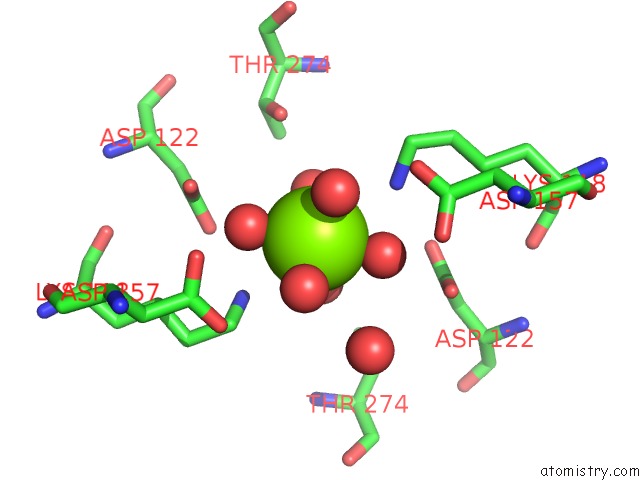
Mono view
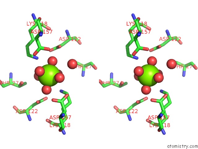
Stereo pair view
Mono view
Stereo pair view
A full contact list of Magnesium with other atoms in the Mg binding
site number 1 of Structural and Functional Study of Succinyl-Ornithine Transaminase From E. Coli within 5.0Å range:
|
Magnesium binding site 2 out of 2 in 4adb
Go back to
Magnesium binding site 2 out
of 2 in the Structural and Functional Study of Succinyl-Ornithine Transaminase From E. Coli
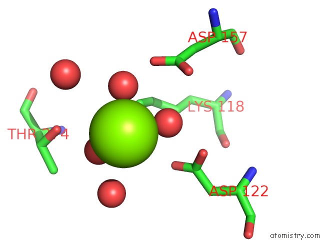
Mono view
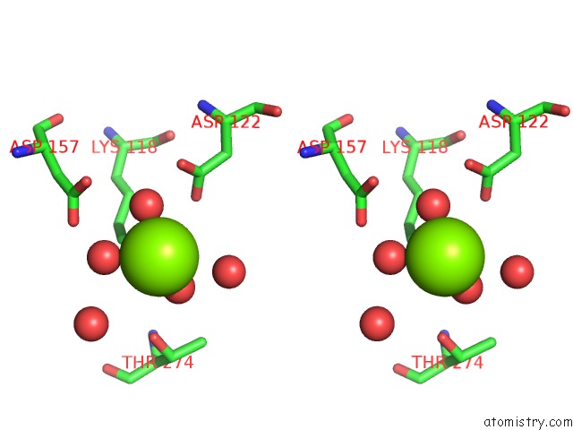
Stereo pair view
Mono view
Stereo pair view
A full contact list of Magnesium with other atoms in the Mg binding
site number 2 of Structural and Functional Study of Succinyl-Ornithine Transaminase From E. Coli within 5.0Å range:
|
Reference:
J.Newman,
S.Seabrook,
R.Surjadi,
C.C.Williams,
D.Lucent,
M.Wilding,
C.Scott,
T.S.Peat.
Determination of the Structure of the Catabolic N- Succinylornithine Transaminase (Astc) From Escherichia Coli. Plos One V. 8 58298 2013.
ISSN: ISSN 1932-6203
PubMed: 23484010
DOI: 10.1371/JOURNAL.PONE.0058298
Page generated: Thu Aug 15 14:30:07 2024
ISSN: ISSN 1932-6203
PubMed: 23484010
DOI: 10.1371/JOURNAL.PONE.0058298
Last articles
Zn in 9MJ5Zn in 9HNW
Zn in 9G0L
Zn in 9FNE
Zn in 9DZN
Zn in 9E0I
Zn in 9D32
Zn in 9DAK
Zn in 8ZXC
Zn in 8ZUF