Magnesium »
PDB 4aau-4ams »
4ajd »
Magnesium in PDB 4ajd: Identification and Structural Characterization of PDE10 Fragment Inhibitors
Enzymatic activity of Identification and Structural Characterization of PDE10 Fragment Inhibitors
All present enzymatic activity of Identification and Structural Characterization of PDE10 Fragment Inhibitors:
3.1.4.17; 3.1.4.35;
3.1.4.17; 3.1.4.35;
Protein crystallography data
The structure of Identification and Structural Characterization of PDE10 Fragment Inhibitors, PDB code: 4ajd
was solved by
P.Johansson,
J.S.Albert,
L.Spadola,
T.Akerud,
E.Back,
P.Hillertz,
R.Horsefeld,
C.Scott,
N.Spear,
G.Tian,
A.Tigerstrom,
D.Aharony,
S.Geschwindner,
with X-Ray Crystallography technique. A brief refinement statistics is given in the table below:
| Resolution Low / High (Å) | 35.00 / 2.30 |
| Space group | P 21 21 21 |
| Cell size a, b, c (Å), α, β, γ (°) | 49.950, 81.490, 158.680, 90.00, 90.00, 90.00 |
| R / Rfree (%) | 18.7 / 24.6 |
Other elements in 4ajd:
The structure of Identification and Structural Characterization of PDE10 Fragment Inhibitors also contains other interesting chemical elements:
| Zinc | (Zn) | 2 atoms |
Magnesium Binding Sites:
The binding sites of Magnesium atom in the Identification and Structural Characterization of PDE10 Fragment Inhibitors
(pdb code 4ajd). This binding sites where shown within
5.0 Angstroms radius around Magnesium atom.
In total 2 binding sites of Magnesium where determined in the Identification and Structural Characterization of PDE10 Fragment Inhibitors, PDB code: 4ajd:
Jump to Magnesium binding site number: 1; 2;
In total 2 binding sites of Magnesium where determined in the Identification and Structural Characterization of PDE10 Fragment Inhibitors, PDB code: 4ajd:
Jump to Magnesium binding site number: 1; 2;
Magnesium binding site 1 out of 2 in 4ajd
Go back to
Magnesium binding site 1 out
of 2 in the Identification and Structural Characterization of PDE10 Fragment Inhibitors
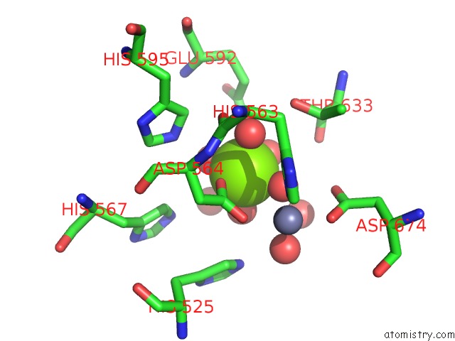
Mono view
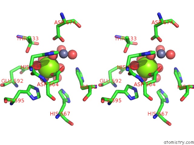
Stereo pair view
Mono view
Stereo pair view
A full contact list of Magnesium with other atoms in the Mg binding
site number 1 of Identification and Structural Characterization of PDE10 Fragment Inhibitors within 5.0Å range:
|
Magnesium binding site 2 out of 2 in 4ajd
Go back to
Magnesium binding site 2 out
of 2 in the Identification and Structural Characterization of PDE10 Fragment Inhibitors
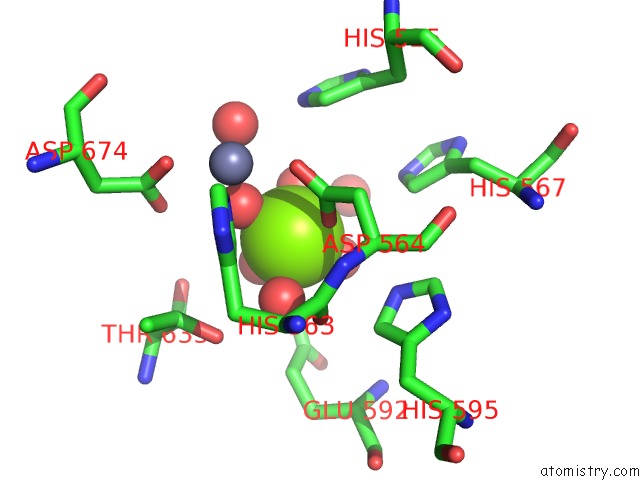
Mono view
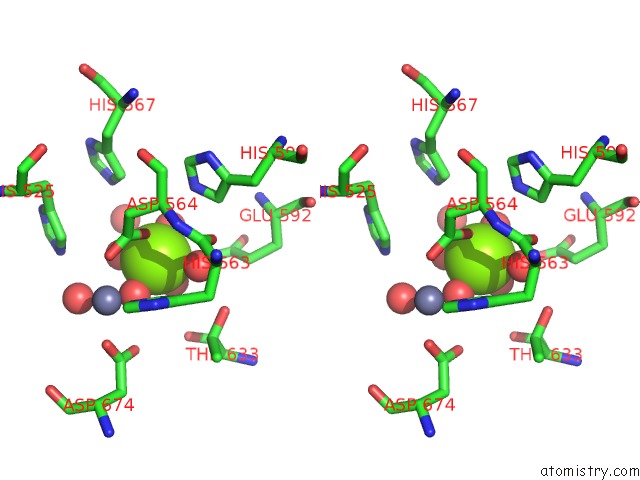
Stereo pair view
Mono view
Stereo pair view
A full contact list of Magnesium with other atoms in the Mg binding
site number 2 of Identification and Structural Characterization of PDE10 Fragment Inhibitors within 5.0Å range:
|
Reference:
P.Johansson,
J.S.Albert,
L.Spadola,
T.Akerud,
E.Back,
P.Hillertz,
R.Horsefeld,
C.Scott,
N.Spear,
G.Tian,
A.Tigerstrom,
D.Aharony,
S.Geschwindner.
Identification and Structural Characterization of PDE10 Fragment Inhibitors To Be Published.
Page generated: Thu Aug 15 14:33:46 2024
Last articles
Ca in 5SB8Ca in 5SB6
Ca in 5SB5
Ca in 5SB3
Ca in 5SB4
Ca in 5S9M
Ca in 5S9N
Ca in 5S9L
Ca in 5S8Q
Ca in 5S8P