Magnesium »
PDB 4aau-4ams »
4ak7 »
Magnesium in PDB 4ak7: Crystal Structure of BPGH117_E303Q in Complex with Neoagarobiose
Protein crystallography data
The structure of Crystal Structure of BPGH117_E303Q in Complex with Neoagarobiose, PDB code: 4ak7
was solved by
J.H.Hehemann,
L.Smyth,
A.Yadav,
D.J.Vocadlo,
A.B.Boraston,
with X-Ray Crystallography technique. A brief refinement statistics is given in the table below:
| Resolution Low / High (Å) | 69.84 / 1.80 |
| Space group | P 21 21 21 |
| Cell size a, b, c (Å), α, β, γ (°) | 83.900, 93.550, 104.960, 90.00, 90.00, 90.00 |
| R / Rfree (%) | 15.544 / 20.854 |
Other elements in 4ak7:
The structure of Crystal Structure of BPGH117_E303Q in Complex with Neoagarobiose also contains other interesting chemical elements:
| Chlorine | (Cl) | 1 atom |
| Calcium | (Ca) | 1 atom |
Magnesium Binding Sites:
The binding sites of Magnesium atom in the Crystal Structure of BPGH117_E303Q in Complex with Neoagarobiose
(pdb code 4ak7). This binding sites where shown within
5.0 Angstroms radius around Magnesium atom.
In total 2 binding sites of Magnesium where determined in the Crystal Structure of BPGH117_E303Q in Complex with Neoagarobiose, PDB code: 4ak7:
Jump to Magnesium binding site number: 1; 2;
In total 2 binding sites of Magnesium where determined in the Crystal Structure of BPGH117_E303Q in Complex with Neoagarobiose, PDB code: 4ak7:
Jump to Magnesium binding site number: 1; 2;
Magnesium binding site 1 out of 2 in 4ak7
Go back to
Magnesium binding site 1 out
of 2 in the Crystal Structure of BPGH117_E303Q in Complex with Neoagarobiose
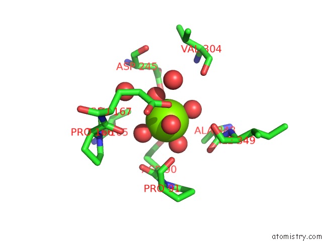
Mono view
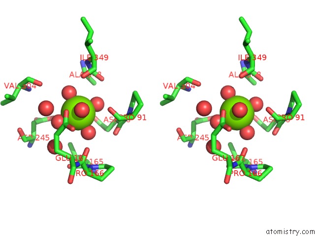
Stereo pair view
Mono view
Stereo pair view
A full contact list of Magnesium with other atoms in the Mg binding
site number 1 of Crystal Structure of BPGH117_E303Q in Complex with Neoagarobiose within 5.0Å range:
|
Magnesium binding site 2 out of 2 in 4ak7
Go back to
Magnesium binding site 2 out
of 2 in the Crystal Structure of BPGH117_E303Q in Complex with Neoagarobiose
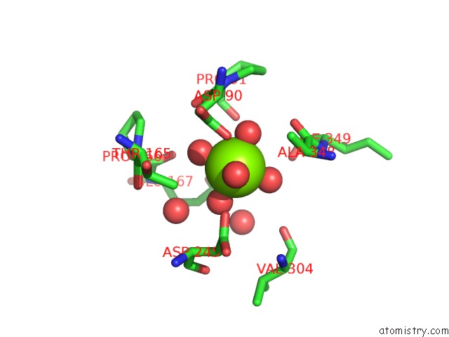
Mono view
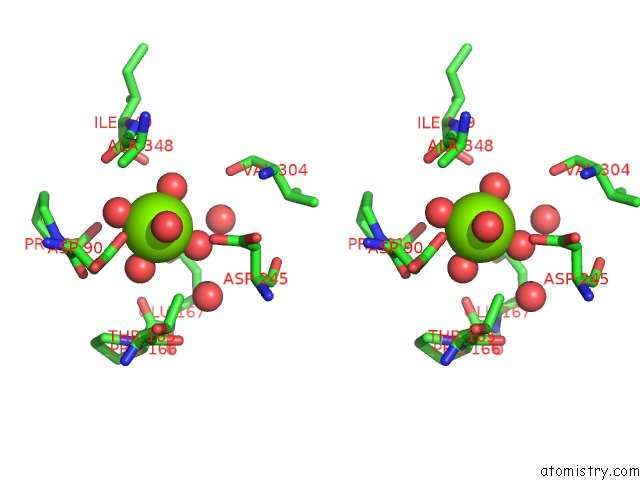
Stereo pair view
Mono view
Stereo pair view
A full contact list of Magnesium with other atoms in the Mg binding
site number 2 of Crystal Structure of BPGH117_E303Q in Complex with Neoagarobiose within 5.0Å range:
|
Reference:
J.H.Hehemann,
L.Smyth,
A.Yadav,
D.J.Vocadlo,
A.B.Boraston.
Analysis of Keystone Enzyme in Agar Hydrolysis Provides Insight Into the Degradation (of A Polysaccharide From) Red Seaweeds. J.Biol.Chem. V. 287 13985 2012.
ISSN: ISSN 0021-9258
PubMed: 22393053
DOI: 10.1074/JBC.M112.345645
Page generated: Thu Aug 15 14:35:11 2024
ISSN: ISSN 0021-9258
PubMed: 22393053
DOI: 10.1074/JBC.M112.345645
Last articles
Fe in 2YXOFe in 2YRS
Fe in 2YXC
Fe in 2YNM
Fe in 2YVJ
Fe in 2YP1
Fe in 2YU2
Fe in 2YU1
Fe in 2YQB
Fe in 2YOO