Magnesium »
PDB 4aas-4amq »
4amk »
Magnesium in PDB 4amk: Fab Fragment of Antiporphrin Antibody 13G10
Protein crystallography data
The structure of Fab Fragment of Antiporphrin Antibody 13G10, PDB code: 4amk
was solved by
J.P.Mahy,
B.Golinelli-Pimpaneau,
with X-Ray Crystallography technique. A brief refinement statistics is given in the table below:
| Resolution Low / High (Å) | 20.0 / 2.05 |
| Space group | C 1 2 1 |
| Cell size a, b, c (Å), α, β, γ (°) | 55.800, 62.750, 113.500, 90.00, 91.65, 90.00 |
| R / Rfree (%) | 22.44 / 27.51 |
Magnesium Binding Sites:
The binding sites of Magnesium atom in the Fab Fragment of Antiporphrin Antibody 13G10
(pdb code 4amk). This binding sites where shown within
5.0 Angstroms radius around Magnesium atom.
In total only one binding site of Magnesium was determined in the Fab Fragment of Antiporphrin Antibody 13G10, PDB code: 4amk:
In total only one binding site of Magnesium was determined in the Fab Fragment of Antiporphrin Antibody 13G10, PDB code: 4amk:
Magnesium binding site 1 out of 1 in 4amk
Go back to
Magnesium binding site 1 out
of 1 in the Fab Fragment of Antiporphrin Antibody 13G10
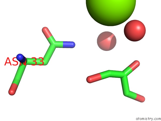
Mono view
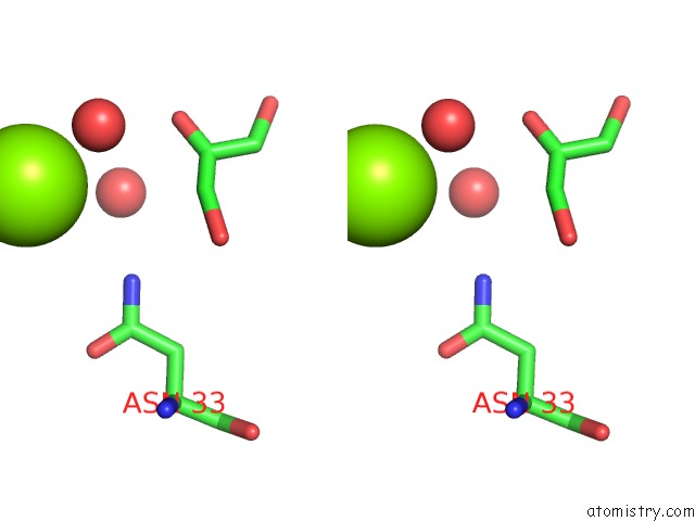
Stereo pair view
Mono view
Stereo pair view
A full contact list of Magnesium with other atoms in the Mg binding
site number 1 of Fab Fragment of Antiporphrin Antibody 13G10 within 5.0Å range:
|
Reference:
V.Munoz Robles,
J.Marechal,
A.Bahloul,
M.Sari,
J.Mahy,
B.Golinelli-Pimpaneau.
Crystal Structure of Two Anti-Porphyrin Antibodies with Peroxidase Activity. Plos One V. 7 51128 2012.
ISSN: ISSN 1932-6203
PubMed: 23240001
DOI: 10.1371/JOURNAL.PONE.0051128
Page generated: Thu Aug 15 14:37:01 2024
ISSN: ISSN 1932-6203
PubMed: 23240001
DOI: 10.1371/JOURNAL.PONE.0051128
Last articles
Zn in 9J0NZn in 9J0O
Zn in 9J0P
Zn in 9FJX
Zn in 9EKB
Zn in 9C0F
Zn in 9CAH
Zn in 9CH0
Zn in 9CH3
Zn in 9CH1