Magnesium »
PDB 4ams-4av6 »
4ap5 »
Magnesium in PDB 4ap5: Crystal Structure of Human POFUT2
Enzymatic activity of Crystal Structure of Human POFUT2
All present enzymatic activity of Crystal Structure of Human POFUT2:
2.4.1.221;
2.4.1.221;
Protein crystallography data
The structure of Crystal Structure of Human POFUT2, PDB code: 4ap5
was solved by
C.Chen,
J.J.Keusch,
D.Klein,
D.Hess,
J.Hofsteenge,
H.Gut,
with X-Ray Crystallography technique. A brief refinement statistics is given in the table below:
| Resolution Low / High (Å) | 29.657 / 3.00 |
| Space group | P 32 2 1 |
| Cell size a, b, c (Å), α, β, γ (°) | 118.628, 118.628, 196.227, 90.00, 90.00, 120.00 |
| R / Rfree (%) | 17.35 / 23.59 |
Other elements in 4ap5:
The structure of Crystal Structure of Human POFUT2 also contains other interesting chemical elements:
| Chlorine | (Cl) | 1 atom |
Magnesium Binding Sites:
The binding sites of Magnesium atom in the Crystal Structure of Human POFUT2
(pdb code 4ap5). This binding sites where shown within
5.0 Angstroms radius around Magnesium atom.
In total only one binding site of Magnesium was determined in the Crystal Structure of Human POFUT2, PDB code: 4ap5:
In total only one binding site of Magnesium was determined in the Crystal Structure of Human POFUT2, PDB code: 4ap5:
Magnesium binding site 1 out of 1 in 4ap5
Go back to
Magnesium binding site 1 out
of 1 in the Crystal Structure of Human POFUT2
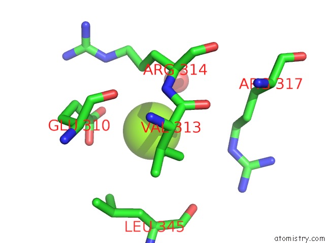
Mono view
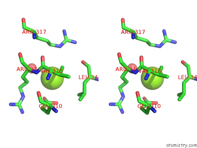
Stereo pair view
Mono view
Stereo pair view
A full contact list of Magnesium with other atoms in the Mg binding
site number 1 of Crystal Structure of Human POFUT2 within 5.0Å range:
|
Reference:
C.I.Chen,
J.J.Keusch,
D.Klein,
D.Hess,
J.Hofsteenge,
H.Gut.
Structure of Human POFUT2: Insights Into Thrombospondin Type 1 Repeat Fold and O-Fucosylation. Embo J. V. 31 3183 2012.
ISSN: ISSN 0261-4189
PubMed: 22588082
DOI: 10.1038/EMBOJ.2012.143
Page generated: Thu Aug 15 14:39:13 2024
ISSN: ISSN 0261-4189
PubMed: 22588082
DOI: 10.1038/EMBOJ.2012.143
Last articles
Zn in 9JYWZn in 9IR4
Zn in 9IR3
Zn in 9GMX
Zn in 9GMW
Zn in 9JEJ
Zn in 9ERF
Zn in 9ERE
Zn in 9EGV
Zn in 9EGW