Magnesium »
PDB 4avq-4b3a »
4b0s »
Magnesium in PDB 4b0s: Structure of the Deamidase-Depupylase Dop of the Prokaryotic Ubiquitin-Like Modification Pathway in Complex with Atp
Protein crystallography data
The structure of Structure of the Deamidase-Depupylase Dop of the Prokaryotic Ubiquitin-Like Modification Pathway in Complex with Atp, PDB code: 4b0s
was solved by
D.Ozcelik,
J.Barandun,
N.Schmitz,
M.Sutter,
E.Guth,
F.F.Damberger,
F.H.-T.Allain,
N.Ban,
E.Weber-Ban,
with X-Ray Crystallography technique. A brief refinement statistics is given in the table below:
| Resolution Low / High (Å) | 47.90 / 2.85 |
| Space group | P 21 21 21 |
| Cell size a, b, c (Å), α, β, γ (°) | 65.360, 70.410, 93.520, 90.00, 90.00, 90.00 |
| R / Rfree (%) | 18.9 / 24.6 |
Magnesium Binding Sites:
The binding sites of Magnesium atom in the Structure of the Deamidase-Depupylase Dop of the Prokaryotic Ubiquitin-Like Modification Pathway in Complex with Atp
(pdb code 4b0s). This binding sites where shown within
5.0 Angstroms radius around Magnesium atom.
In total 2 binding sites of Magnesium where determined in the Structure of the Deamidase-Depupylase Dop of the Prokaryotic Ubiquitin-Like Modification Pathway in Complex with Atp, PDB code: 4b0s:
Jump to Magnesium binding site number: 1; 2;
In total 2 binding sites of Magnesium where determined in the Structure of the Deamidase-Depupylase Dop of the Prokaryotic Ubiquitin-Like Modification Pathway in Complex with Atp, PDB code: 4b0s:
Jump to Magnesium binding site number: 1; 2;
Magnesium binding site 1 out of 2 in 4b0s
Go back to
Magnesium binding site 1 out
of 2 in the Structure of the Deamidase-Depupylase Dop of the Prokaryotic Ubiquitin-Like Modification Pathway in Complex with Atp
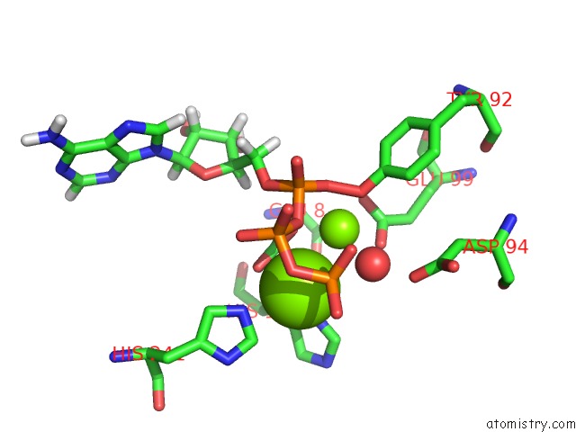
Mono view
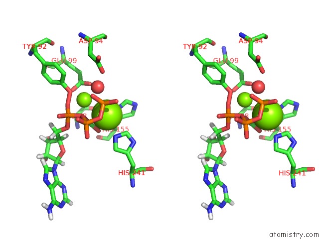
Stereo pair view
Mono view
Stereo pair view
A full contact list of Magnesium with other atoms in the Mg binding
site number 1 of Structure of the Deamidase-Depupylase Dop of the Prokaryotic Ubiquitin-Like Modification Pathway in Complex with Atp within 5.0Å range:
|
Magnesium binding site 2 out of 2 in 4b0s
Go back to
Magnesium binding site 2 out
of 2 in the Structure of the Deamidase-Depupylase Dop of the Prokaryotic Ubiquitin-Like Modification Pathway in Complex with Atp
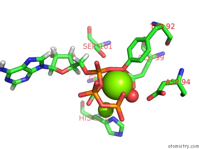
Mono view
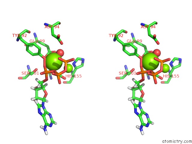
Stereo pair view
Mono view
Stereo pair view
A full contact list of Magnesium with other atoms in the Mg binding
site number 2 of Structure of the Deamidase-Depupylase Dop of the Prokaryotic Ubiquitin-Like Modification Pathway in Complex with Atp within 5.0Å range:
|
Reference:
D.Ozcelik,
J.Barandun,
N.Schmitz,
M.Sutter,
E.Guth,
F.F.Damberger,
F.H.-T.Allain,
N.Ban,
E.Weber-Ban.
Structures of Pup Ligase Pafa and Depupylase Dop From the Prokaryotic Ubiquitin-Like Modification Pathway. Nat.Commun. V. 3 1014 2012.
ISSN: ESSN 2041-1723
PubMed: 22910360
DOI: 10.1038/NCOMMS2009
Page generated: Mon Aug 11 06:06:59 2025
ISSN: ESSN 2041-1723
PubMed: 22910360
DOI: 10.1038/NCOMMS2009
Last articles
Mg in 4DR3Mg in 4DR1
Mg in 4DPG
Mg in 4DQP
Mg in 4DQQ
Mg in 4DPM
Mg in 4DPV
Mg in 4DQI
Mg in 4DOB
Mg in 4DOC