Magnesium »
PDB 4avq-4b3a »
4b1w »
Magnesium in PDB 4b1w: Structure of the PHACTR1 Rpel-2 Domain Bound to Actin
Protein crystallography data
The structure of Structure of the PHACTR1 Rpel-2 Domain Bound to Actin, PDB code: 4b1w
was solved by
S.Mouilleron,
M.Wiezlak,
N.O'reilly,
R.Treisman,
N.Q.Mcdonald,
with X-Ray Crystallography technique. A brief refinement statistics is given in the table below:
| Resolution Low / High (Å) | 31.73 / 1.95 |
| Space group | I 4 2 2 |
| Cell size a, b, c (Å), α, β, γ (°) | 128.220, 128.220, 135.470, 90.00, 90.00, 90.00 |
| R / Rfree (%) | 17.9 / 21.3 |
Magnesium Binding Sites:
The binding sites of Magnesium atom in the Structure of the PHACTR1 Rpel-2 Domain Bound to Actin
(pdb code 4b1w). This binding sites where shown within
5.0 Angstroms radius around Magnesium atom.
In total only one binding site of Magnesium was determined in the Structure of the PHACTR1 Rpel-2 Domain Bound to Actin, PDB code: 4b1w:
In total only one binding site of Magnesium was determined in the Structure of the PHACTR1 Rpel-2 Domain Bound to Actin, PDB code: 4b1w:
Magnesium binding site 1 out of 1 in 4b1w
Go back to
Magnesium binding site 1 out
of 1 in the Structure of the PHACTR1 Rpel-2 Domain Bound to Actin
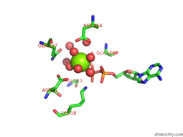
Mono view
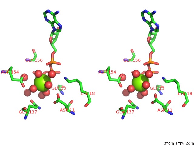
Stereo pair view
Mono view
Stereo pair view
A full contact list of Magnesium with other atoms in the Mg binding
site number 1 of Structure of the PHACTR1 Rpel-2 Domain Bound to Actin within 5.0Å range:
|
Reference:
S.Mouilleron,
M.Wiezlak,
N.O'reilly,
R.Treisman,
N.Q.Mcdonald.
Structures of the PHACTR1 Rpel Domain and Rpel Motif Complexes with G-Actin Reveal the Molecular Basis For Actin Binding Cooperativity. Structure V. 20 1960 2012.
ISSN: ISSN 1878-4186
PubMed: 23041370
DOI: 10.1016/J.STR.2012.08.031
Page generated: Mon Aug 11 06:08:17 2025
ISSN: ISSN 1878-4186
PubMed: 23041370
DOI: 10.1016/J.STR.2012.08.031
Last articles
Mg in 4DUXMg in 4DUW
Mg in 4DUV
Mg in 4DUO
Mg in 4DUG
Mg in 4DTY
Mg in 4DTW
Mg in 4DTH
Mg in 4DTF
Mg in 4DSU