Magnesium »
PDB 4avq-4b3a »
4b20 »
Magnesium in PDB 4b20: Structural Basis of Dna Loop Recognition By Endonuclease V
Enzymatic activity of Structural Basis of Dna Loop Recognition By Endonuclease V
All present enzymatic activity of Structural Basis of Dna Loop Recognition By Endonuclease V:
3.1.21.7;
3.1.21.7;
Protein crystallography data
The structure of Structural Basis of Dna Loop Recognition By Endonuclease V, PDB code: 4b20
was solved by
I.Rosnes,
A.D.Rowe,
R.J.Forstrom,
I.Alseth,
M.Bjoras,
B.Dalhus,
with X-Ray Crystallography technique. A brief refinement statistics is given in the table below:
| Resolution Low / High (Å) | 60.0 / 2.75 |
| Space group | I 2 2 2 |
| Cell size a, b, c (Å), α, β, γ (°) | 55.231, 135.370, 194.993, 90.00, 90.00, 90.00 |
| R / Rfree (%) | 22.74 / 29.06 |
Magnesium Binding Sites:
The binding sites of Magnesium atom in the Structural Basis of Dna Loop Recognition By Endonuclease V
(pdb code 4b20). This binding sites where shown within
5.0 Angstroms radius around Magnesium atom.
In total 2 binding sites of Magnesium where determined in the Structural Basis of Dna Loop Recognition By Endonuclease V, PDB code: 4b20:
Jump to Magnesium binding site number: 1; 2;
In total 2 binding sites of Magnesium where determined in the Structural Basis of Dna Loop Recognition By Endonuclease V, PDB code: 4b20:
Jump to Magnesium binding site number: 1; 2;
Magnesium binding site 1 out of 2 in 4b20
Go back to
Magnesium binding site 1 out
of 2 in the Structural Basis of Dna Loop Recognition By Endonuclease V

Mono view
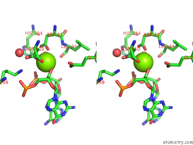
Stereo pair view
Mono view
Stereo pair view
A full contact list of Magnesium with other atoms in the Mg binding
site number 1 of Structural Basis of Dna Loop Recognition By Endonuclease V within 5.0Å range:
|
Magnesium binding site 2 out of 2 in 4b20
Go back to
Magnesium binding site 2 out
of 2 in the Structural Basis of Dna Loop Recognition By Endonuclease V
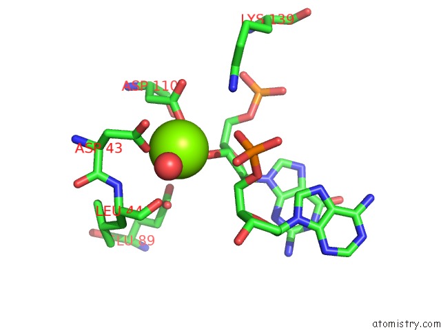
Mono view

Stereo pair view
Mono view
Stereo pair view
A full contact list of Magnesium with other atoms in the Mg binding
site number 2 of Structural Basis of Dna Loop Recognition By Endonuclease V within 5.0Å range:
|
Reference:
I.Rosnes,
A.D.Rowe,
E.S.Vik,
R.J.Forstrom,
I.Alseth,
M.Bjoras,
B.Dalhus.
Structural Basis of Dna Loop Recognition By Endonuclease V. Structure V. 21 257 2013.
ISSN: ISSN 0969-2126
PubMed: 23313664
DOI: 10.1016/J.STR.2012.12.007
Page generated: Mon Aug 11 06:08:35 2025
ISSN: ISSN 0969-2126
PubMed: 23313664
DOI: 10.1016/J.STR.2012.12.007
Last articles
Mg in 4DUXMg in 4DUW
Mg in 4DUV
Mg in 4DUO
Mg in 4DUG
Mg in 4DTY
Mg in 4DTW
Mg in 4DTH
Mg in 4DTF
Mg in 4DSU