Magnesium »
PDB 4beb-4brq »
4bjr »
Magnesium in PDB 4bjr: Crystal Structure of the Complex Between Prokaryotic Ubiquitin-Like Protein Pup and Its Ligase Pafa
Protein crystallography data
The structure of Crystal Structure of the Complex Between Prokaryotic Ubiquitin-Like Protein Pup and Its Ligase Pafa, PDB code: 4bjr
was solved by
J.Barandun,
C.L.Delley,
N.Ban,
E.Weber-Ban,
with X-Ray Crystallography technique. A brief refinement statistics is given in the table below:
| Resolution Low / High (Å) | 39.131 / 2.80 |
| Space group | P 21 21 21 |
| Cell size a, b, c (Å), α, β, γ (°) | 63.840, 84.020, 215.110, 90.00, 90.00, 90.00 |
| R / Rfree (%) | 21.31 / 24.75 |
Magnesium Binding Sites:
The binding sites of Magnesium atom in the Crystal Structure of the Complex Between Prokaryotic Ubiquitin-Like Protein Pup and Its Ligase Pafa
(pdb code 4bjr). This binding sites where shown within
5.0 Angstroms radius around Magnesium atom.
In total 3 binding sites of Magnesium where determined in the Crystal Structure of the Complex Between Prokaryotic Ubiquitin-Like Protein Pup and Its Ligase Pafa, PDB code: 4bjr:
Jump to Magnesium binding site number: 1; 2; 3;
In total 3 binding sites of Magnesium where determined in the Crystal Structure of the Complex Between Prokaryotic Ubiquitin-Like Protein Pup and Its Ligase Pafa, PDB code: 4bjr:
Jump to Magnesium binding site number: 1; 2; 3;
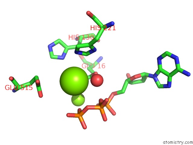
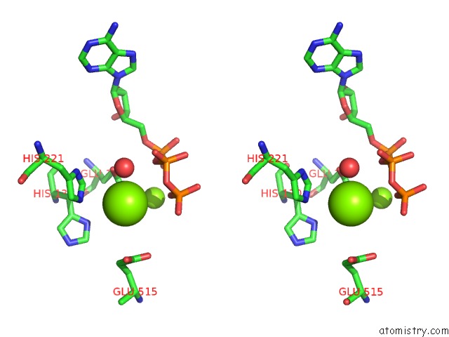
Magnesium binding site 1 out of 3 in 4bjr
Go back to
Magnesium binding site 1 out
of 3 in the Crystal Structure of the Complex Between Prokaryotic Ubiquitin-Like Protein Pup and Its Ligase Pafa
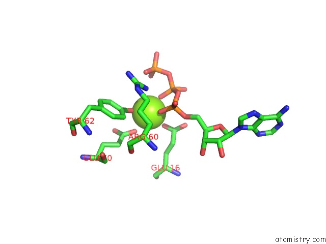
Mono view
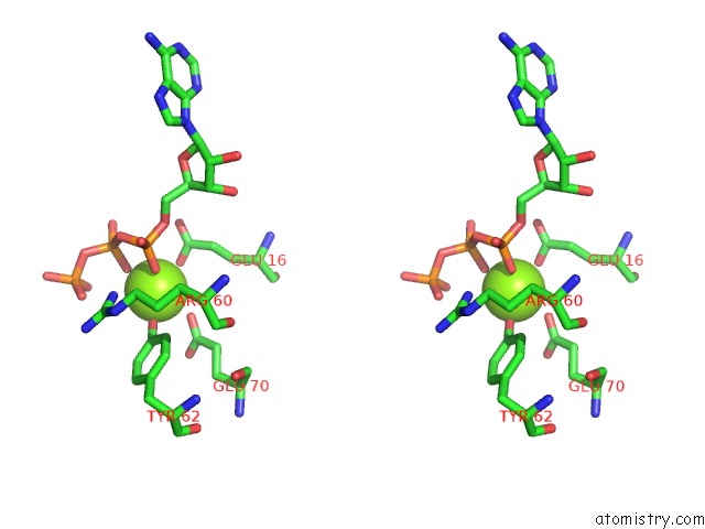
Stereo pair view
Mono view
Stereo pair view
A full contact list of Magnesium with other atoms in the Mg binding
site number 1 of Crystal Structure of the Complex Between Prokaryotic Ubiquitin-Like Protein Pup and Its Ligase Pafa within 5.0Å range:
|
Magnesium binding site 2 out of 3 in 4bjr
Go back to
Magnesium binding site 2 out
of 3 in the Crystal Structure of the Complex Between Prokaryotic Ubiquitin-Like Protein Pup and Its Ligase Pafa
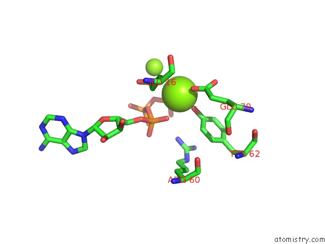
Mono view
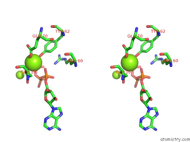
Stereo pair view
Mono view
Stereo pair view
A full contact list of Magnesium with other atoms in the Mg binding
site number 2 of Crystal Structure of the Complex Between Prokaryotic Ubiquitin-Like Protein Pup and Its Ligase Pafa within 5.0Å range:
|
Magnesium binding site 3 out of 3 in 4bjr
Go back to
Magnesium binding site 3 out
of 3 in the Crystal Structure of the Complex Between Prokaryotic Ubiquitin-Like Protein Pup and Its Ligase Pafa
Mono view
Stereo pair view
Mono view
Stereo pair view
A full contact list of Magnesium with other atoms in the Mg binding
site number 3 of Crystal Structure of the Complex Between Prokaryotic Ubiquitin-Like Protein Pup and Its Ligase Pafa within 5.0Å range:
|
Reference:
J.Barandun,
C.L.Delley,
N.Ban,
E.Weber-Ban.
Crystal Structure of the Complex Between Prokaryotic Ubiquitin-Like Protein Pup and Its Ligase Pafa. J.Am.Chem.Soc. V. 135 6794 2013.
ISSN: ISSN 0002-7863
PubMed: 23601177
DOI: 10.1021/JA4024012
Page generated: Thu Aug 15 16:29:17 2024
ISSN: ISSN 0002-7863
PubMed: 23601177
DOI: 10.1021/JA4024012
Last articles
Fe in 2YXOFe in 2YRS
Fe in 2YXC
Fe in 2YNM
Fe in 2YVJ
Fe in 2YP1
Fe in 2YU2
Fe in 2YU1
Fe in 2YQB
Fe in 2YOO