Magnesium »
PDB 4beb-4brq »
4brl »
Magnesium in PDB 4brl: Legionella Pneumophila NTPDASE1 Crystal Form III (Closed) in Complex with Transition State Mimic Guanosine 5'-Phosphovanadate
Enzymatic activity of Legionella Pneumophila NTPDASE1 Crystal Form III (Closed) in Complex with Transition State Mimic Guanosine 5'-Phosphovanadate
All present enzymatic activity of Legionella Pneumophila NTPDASE1 Crystal Form III (Closed) in Complex with Transition State Mimic Guanosine 5'-Phosphovanadate:
3.6.1.5;
3.6.1.5;
Protein crystallography data
The structure of Legionella Pneumophila NTPDASE1 Crystal Form III (Closed) in Complex with Transition State Mimic Guanosine 5'-Phosphovanadate, PDB code: 4brl
was solved by
M.Zebisch,
P.Schaefer,
P.Lauble,
N.Straeter,
with X-Ray Crystallography technique. A brief refinement statistics is given in the table below:
| Resolution Low / High (Å) | 19.88 / 1.60 |
| Space group | P 21 21 21 |
| Cell size a, b, c (Å), α, β, γ (°) | 80.994, 83.463, 109.289, 90.00, 90.00, 90.00 |
| R / Rfree (%) | 14.416 / 19.773 |
Other elements in 4brl:
The structure of Legionella Pneumophila NTPDASE1 Crystal Form III (Closed) in Complex with Transition State Mimic Guanosine 5'-Phosphovanadate also contains other interesting chemical elements:
| Chlorine | (Cl) | 3 atoms |
| Vanadium | (V) | 2 atoms |
Magnesium Binding Sites:
The binding sites of Magnesium atom in the Legionella Pneumophila NTPDASE1 Crystal Form III (Closed) in Complex with Transition State Mimic Guanosine 5'-Phosphovanadate
(pdb code 4brl). This binding sites where shown within
5.0 Angstroms radius around Magnesium atom.
In total 2 binding sites of Magnesium where determined in the Legionella Pneumophila NTPDASE1 Crystal Form III (Closed) in Complex with Transition State Mimic Guanosine 5'-Phosphovanadate, PDB code: 4brl:
Jump to Magnesium binding site number: 1; 2;
In total 2 binding sites of Magnesium where determined in the Legionella Pneumophila NTPDASE1 Crystal Form III (Closed) in Complex with Transition State Mimic Guanosine 5'-Phosphovanadate, PDB code: 4brl:
Jump to Magnesium binding site number: 1; 2;
Magnesium binding site 1 out of 2 in 4brl
Go back to
Magnesium binding site 1 out
of 2 in the Legionella Pneumophila NTPDASE1 Crystal Form III (Closed) in Complex with Transition State Mimic Guanosine 5'-Phosphovanadate

Mono view
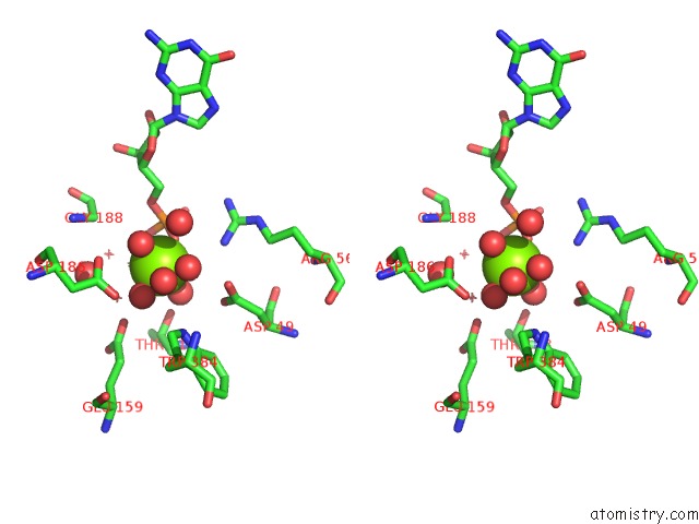
Stereo pair view
Mono view
Stereo pair view
A full contact list of Magnesium with other atoms in the Mg binding
site number 1 of Legionella Pneumophila NTPDASE1 Crystal Form III (Closed) in Complex with Transition State Mimic Guanosine 5'-Phosphovanadate within 5.0Å range:
|
Magnesium binding site 2 out of 2 in 4brl
Go back to
Magnesium binding site 2 out
of 2 in the Legionella Pneumophila NTPDASE1 Crystal Form III (Closed) in Complex with Transition State Mimic Guanosine 5'-Phosphovanadate
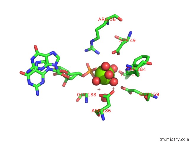
Mono view

Stereo pair view
Mono view
Stereo pair view
A full contact list of Magnesium with other atoms in the Mg binding
site number 2 of Legionella Pneumophila NTPDASE1 Crystal Form III (Closed) in Complex with Transition State Mimic Guanosine 5'-Phosphovanadate within 5.0Å range:
|
Reference:
M.Zebisch,
M.Krauss,
P.Schaefer,
P.Lauble,
N.Straeter.
Crystallographic Snapshots Along the Reaction Pathway of Nucleoside Triphosphate Diphosphohydrolases Structure V. 21 1460 2013.
ISSN: ISSN 0969-2126
PubMed: 23830739
DOI: 10.1016/J.STR.2013.05.016
Page generated: Thu Aug 15 16:32:52 2024
ISSN: ISSN 0969-2126
PubMed: 23830739
DOI: 10.1016/J.STR.2013.05.016
Last articles
Cl in 5WO3Cl in 5WO4
Cl in 5WPN
Cl in 5WO2
Cl in 5WO1
Cl in 5WNY
Cl in 5WO0
Cl in 5WNW
Cl in 5WNZ
Cl in 5WNX