Magnesium »
PDB 4brs-4c2z »
4bvs »
Magnesium in PDB 4bvs: Cyanuric Acid Hydrolase: Evolutionary Innovation By Structural Concatenation.
Enzymatic activity of Cyanuric Acid Hydrolase: Evolutionary Innovation By Structural Concatenation.
All present enzymatic activity of Cyanuric Acid Hydrolase: Evolutionary Innovation By Structural Concatenation.:
3.5.2.15;
3.5.2.15;
Protein crystallography data
The structure of Cyanuric Acid Hydrolase: Evolutionary Innovation By Structural Concatenation., PDB code: 4bvs
was solved by
T.S.Peat,
S.Balotra,
M.Wilding,
N.G.French,
L.J.Briggs,
S.Panjikar,
N.Cowieson,
J.Newman,
C.Scott,
with X-Ray Crystallography technique. A brief refinement statistics is given in the table below:
| Resolution Low / High (Å) | 42.29 / 2.60 |
| Space group | H 3 2 |
| Cell size a, b, c (Å), α, β, γ (°) | 128.365, 128.365, 228.412, 90.00, 90.00, 120.00 |
| R / Rfree (%) | 17.663 / 21.572 |
Magnesium Binding Sites:
The binding sites of Magnesium atom in the Cyanuric Acid Hydrolase: Evolutionary Innovation By Structural Concatenation.
(pdb code 4bvs). This binding sites where shown within
5.0 Angstroms radius around Magnesium atom.
In total 2 binding sites of Magnesium where determined in the Cyanuric Acid Hydrolase: Evolutionary Innovation By Structural Concatenation., PDB code: 4bvs:
Jump to Magnesium binding site number: 1; 2;
In total 2 binding sites of Magnesium where determined in the Cyanuric Acid Hydrolase: Evolutionary Innovation By Structural Concatenation., PDB code: 4bvs:
Jump to Magnesium binding site number: 1; 2;
Magnesium binding site 1 out of 2 in 4bvs
Go back to
Magnesium binding site 1 out
of 2 in the Cyanuric Acid Hydrolase: Evolutionary Innovation By Structural Concatenation.
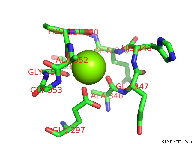
Mono view
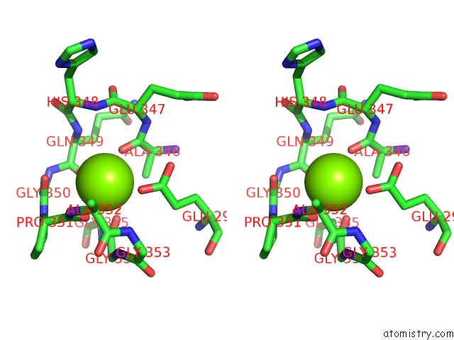
Stereo pair view
Mono view
Stereo pair view
A full contact list of Magnesium with other atoms in the Mg binding
site number 1 of Cyanuric Acid Hydrolase: Evolutionary Innovation By Structural Concatenation. within 5.0Å range:
|
Magnesium binding site 2 out of 2 in 4bvs
Go back to
Magnesium binding site 2 out
of 2 in the Cyanuric Acid Hydrolase: Evolutionary Innovation By Structural Concatenation.
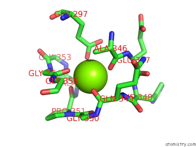
Mono view
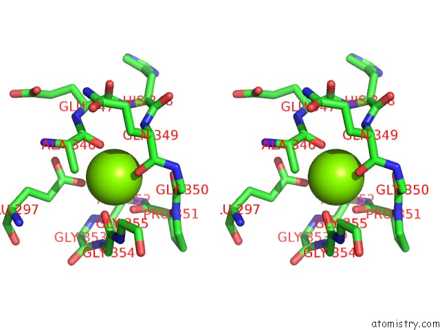
Stereo pair view
Mono view
Stereo pair view
A full contact list of Magnesium with other atoms in the Mg binding
site number 2 of Cyanuric Acid Hydrolase: Evolutionary Innovation By Structural Concatenation. within 5.0Å range:
|
Reference:
T.S.Peat,
S.Balotra,
M.Wilding,
N.G.French,
L.J.Briggs,
S.Panjikar,
N.Cowieson,
J.Newman,
C.Scott.
Cyanuric Acid Hydrolase: Evolutionary Innovation By Structural Concatenation. Mol.Microbiol. V. 88 1149 2013.
ISSN: ISSN 0950-382X
PubMed: 23651355
DOI: 10.1111/MMI.12249
Page generated: Mon Aug 11 07:05:42 2025
ISSN: ISSN 0950-382X
PubMed: 23651355
DOI: 10.1111/MMI.12249
Last articles
Mg in 5BTFMg in 5BTD
Mg in 5BTC
Mg in 5BTA
Mg in 5BON
Mg in 5BSU
Mg in 5BST
Mg in 5BSM
Mg in 5BSK
Mg in 5BS8