Magnesium »
PDB 4brq-4c2y »
4bwx »
Magnesium in PDB 4bwx: Structure of Neurospora Crassa PAN3 Pseudokinase Mutant
Protein crystallography data
The structure of Structure of Neurospora Crassa PAN3 Pseudokinase Mutant, PDB code: 4bwx
was solved by
M.Christie,
A.Boland,
E.Huntzinger,
O.Weichenrieder,
E.Izaurralde,
with X-Ray Crystallography technique. A brief refinement statistics is given in the table below:
| Resolution Low / High (Å) | 45.156 / 2.85 |
| Space group | P 65 |
| Cell size a, b, c (Å), α, β, γ (°) | 90.312, 90.312, 230.609, 90.00, 90.00, 120.00 |
| R / Rfree (%) | 21.1 / 24.24 |
Magnesium Binding Sites:
The binding sites of Magnesium atom in the Structure of Neurospora Crassa PAN3 Pseudokinase Mutant
(pdb code 4bwx). This binding sites where shown within
5.0 Angstroms radius around Magnesium atom.
In total 2 binding sites of Magnesium where determined in the Structure of Neurospora Crassa PAN3 Pseudokinase Mutant, PDB code: 4bwx:
Jump to Magnesium binding site number: 1; 2;
In total 2 binding sites of Magnesium where determined in the Structure of Neurospora Crassa PAN3 Pseudokinase Mutant, PDB code: 4bwx:
Jump to Magnesium binding site number: 1; 2;
Magnesium binding site 1 out of 2 in 4bwx
Go back to
Magnesium binding site 1 out
of 2 in the Structure of Neurospora Crassa PAN3 Pseudokinase Mutant
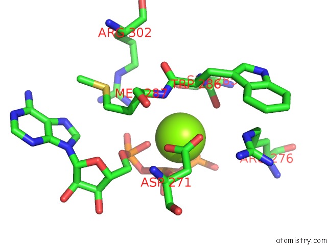
Mono view
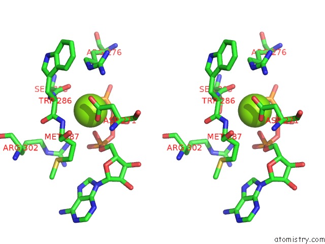
Stereo pair view
Mono view
Stereo pair view
A full contact list of Magnesium with other atoms in the Mg binding
site number 1 of Structure of Neurospora Crassa PAN3 Pseudokinase Mutant within 5.0Å range:
|
Magnesium binding site 2 out of 2 in 4bwx
Go back to
Magnesium binding site 2 out
of 2 in the Structure of Neurospora Crassa PAN3 Pseudokinase Mutant
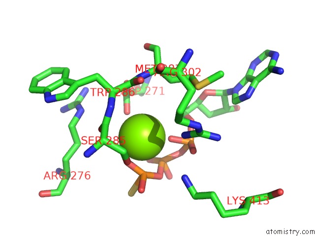
Mono view
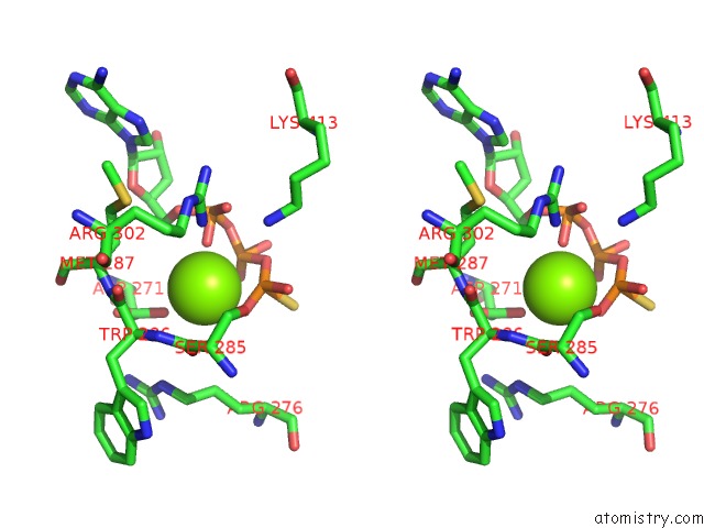
Stereo pair view
Mono view
Stereo pair view
A full contact list of Magnesium with other atoms in the Mg binding
site number 2 of Structure of Neurospora Crassa PAN3 Pseudokinase Mutant within 5.0Å range:
|
Reference:
M.Christie,
A.Boland,
E.Huntzinger,
O.Weichenrieder,
E.Izaurralde.
Structure of the PAN3 Pseudokinase Reveals the Basis For Interactions with the PAN2 Deadenylase and the GW182 Proteins Mol.Cell V. 51 360 2013.
ISSN: ISSN 1097-2765
PubMed: 23932717
DOI: 10.1016/J.MOLCEL.2013.07.011
Page generated: Thu Aug 15 16:36:17 2024
ISSN: ISSN 1097-2765
PubMed: 23932717
DOI: 10.1016/J.MOLCEL.2013.07.011
Last articles
Zn in 9J0NZn in 9J0O
Zn in 9J0P
Zn in 9FJX
Zn in 9EKB
Zn in 9C0F
Zn in 9CAH
Zn in 9CH0
Zn in 9CH3
Zn in 9CH1