Magnesium »
PDB 4brq-4c2y »
4bx3 »
Magnesium in PDB 4bx3: Crystal Structure of Murine Chronophin (Pyridoxal Phosphate Phosphatase)
Enzymatic activity of Crystal Structure of Murine Chronophin (Pyridoxal Phosphate Phosphatase)
All present enzymatic activity of Crystal Structure of Murine Chronophin (Pyridoxal Phosphate Phosphatase):
3.1.3.3; 3.1.3.74;
3.1.3.3; 3.1.3.74;
Protein crystallography data
The structure of Crystal Structure of Murine Chronophin (Pyridoxal Phosphate Phosphatase), PDB code: 4bx3
was solved by
G.Knobloch,
A.Gohla,
H.Schindelin,
with X-Ray Crystallography technique. A brief refinement statistics is given in the table below:
| Resolution Low / High (Å) | 44.659 / 2.19 |
| Space group | I 2 3 |
| Cell size a, b, c (Å), α, β, γ (°) | 167.100, 167.100, 167.100, 90.00, 90.00, 90.00 |
| R / Rfree (%) | 17.07 / 20.67 |
Magnesium Binding Sites:
The binding sites of Magnesium atom in the Crystal Structure of Murine Chronophin (Pyridoxal Phosphate Phosphatase)
(pdb code 4bx3). This binding sites where shown within
5.0 Angstroms radius around Magnesium atom.
In total 2 binding sites of Magnesium where determined in the Crystal Structure of Murine Chronophin (Pyridoxal Phosphate Phosphatase), PDB code: 4bx3:
Jump to Magnesium binding site number: 1; 2;
In total 2 binding sites of Magnesium where determined in the Crystal Structure of Murine Chronophin (Pyridoxal Phosphate Phosphatase), PDB code: 4bx3:
Jump to Magnesium binding site number: 1; 2;
Magnesium binding site 1 out of 2 in 4bx3
Go back to
Magnesium binding site 1 out
of 2 in the Crystal Structure of Murine Chronophin (Pyridoxal Phosphate Phosphatase)
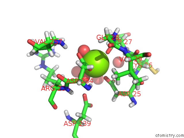
Mono view
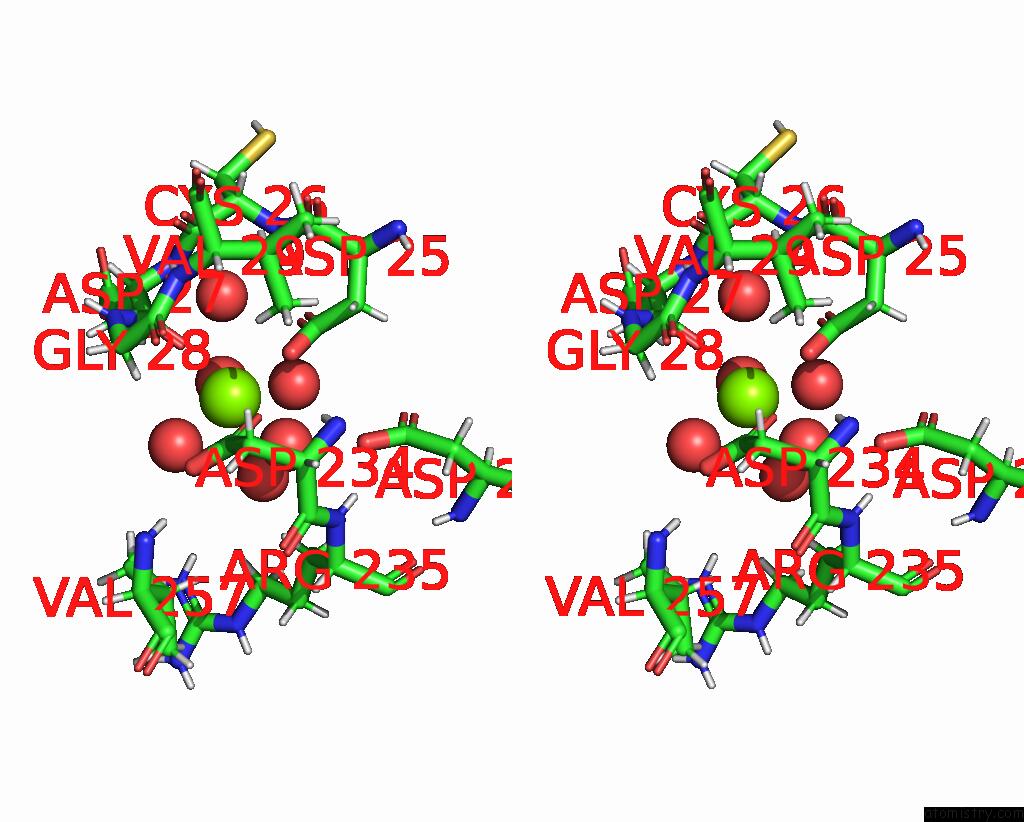
Stereo pair view
Mono view
Stereo pair view
A full contact list of Magnesium with other atoms in the Mg binding
site number 1 of Crystal Structure of Murine Chronophin (Pyridoxal Phosphate Phosphatase) within 5.0Å range:
|
Magnesium binding site 2 out of 2 in 4bx3
Go back to
Magnesium binding site 2 out
of 2 in the Crystal Structure of Murine Chronophin (Pyridoxal Phosphate Phosphatase)
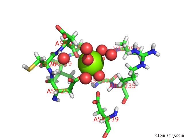
Mono view
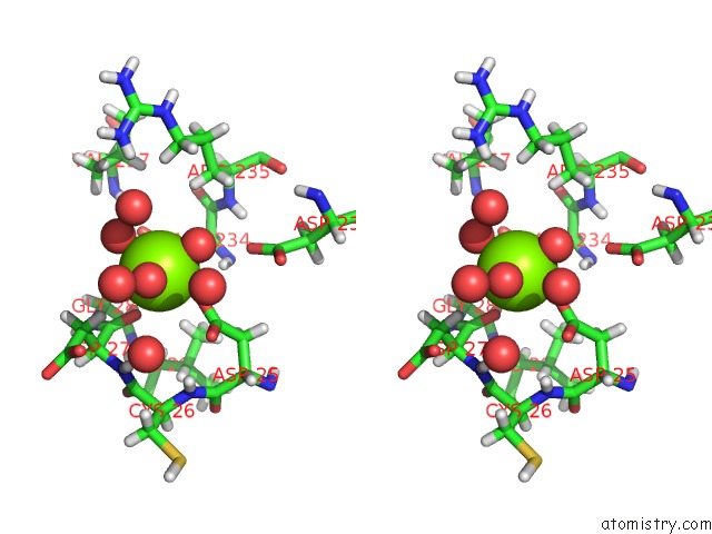
Stereo pair view
Mono view
Stereo pair view
A full contact list of Magnesium with other atoms in the Mg binding
site number 2 of Crystal Structure of Murine Chronophin (Pyridoxal Phosphate Phosphatase) within 5.0Å range:
|
Reference:
C.Kestler,
G.Knobloch,
I.Tessmer,
E.Jeanclos,
H.Schindelin,
A.Gohla.
Chronophin Dimerization Is Required For Proper Positioning of Its Substrate Specificity Loop. J.Biol.Chem. V. 289 3094 2014.
ISSN: ISSN 0021-9258
PubMed: 24338687
DOI: 10.1074/JBC.M113.536482
Page generated: Thu Aug 15 16:36:35 2024
ISSN: ISSN 0021-9258
PubMed: 24338687
DOI: 10.1074/JBC.M113.536482
Last articles
Zn in 9J0NZn in 9J0O
Zn in 9J0P
Zn in 9FJX
Zn in 9EKB
Zn in 9C0F
Zn in 9CAH
Zn in 9CH0
Zn in 9CH3
Zn in 9CH1