Magnesium in PDB 4c5c: The X-Ray Crystal Structure of D-Alanyl-D-Alanine Ligase in Complex with Adp and D-Ala-D-Ala
Enzymatic activity of The X-Ray Crystal Structure of D-Alanyl-D-Alanine Ligase in Complex with Adp and D-Ala-D-Ala
All present enzymatic activity of The X-Ray Crystal Structure of D-Alanyl-D-Alanine Ligase in Complex with Adp and D-Ala-D-Ala:
6.3.2.4;
Protein crystallography data
The structure of The X-Ray Crystal Structure of D-Alanyl-D-Alanine Ligase in Complex with Adp and D-Ala-D-Ala, PDB code: 4c5c
was solved by
S.Batson,
V.Majce,
A.J.Lloyd,
D.Rea,
C.W.G.Fishwick,
K.J.Simmons,
V.Fulop,
D.I.Roper,
with X-Ray Crystallography technique. A brief refinement statistics is given in the table below:
Resolution Low / High (Å)
|
48.90 /
1.40
|
Space group
|
P 21 21 21
|
Cell size a, b, c (Å), α, β, γ (°)
|
51.230,
97.800,
110.120,
90.00,
90.00,
90.00
|
R / Rfree (%)
|
17.975 /
20.341
|
Magnesium Binding Sites:
The binding sites of Magnesium atom in the The X-Ray Crystal Structure of D-Alanyl-D-Alanine Ligase in Complex with Adp and D-Ala-D-Ala
(pdb code 4c5c). This binding sites where shown within
5.0 Angstroms radius around Magnesium atom.
In total 7 binding sites of Magnesium where determined in the
The X-Ray Crystal Structure of D-Alanyl-D-Alanine Ligase in Complex with Adp and D-Ala-D-Ala, PDB code: 4c5c:
Jump to Magnesium binding site number:
1;
2;
3;
4;
5;
6;
7;
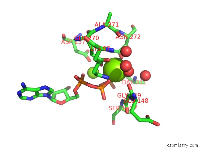
Magnesium binding site 1 out
of 7 in 4c5c
Go back to
Magnesium Binding Sites List in 4c5c
Magnesium binding site 1 out
of 7 in the The X-Ray Crystal Structure of D-Alanyl-D-Alanine Ligase in Complex with Adp and D-Ala-D-Ala
Mono view
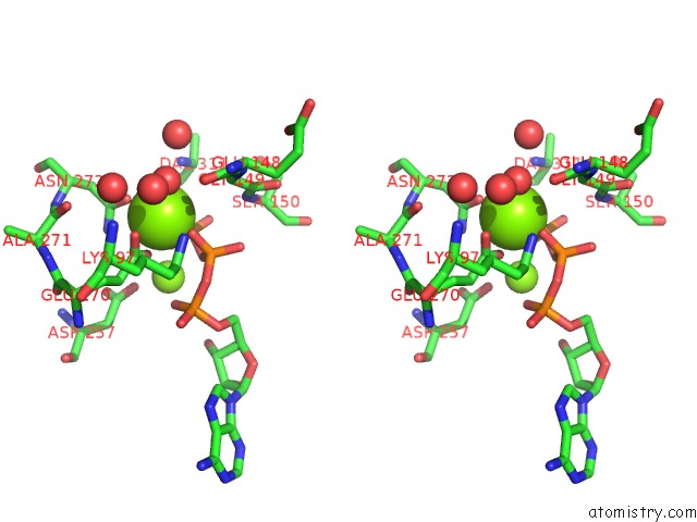
Stereo pair view
|
A full contact list of Magnesium with other atoms in the Mg binding
site number 1 of The X-Ray Crystal Structure of D-Alanyl-D-Alanine Ligase in Complex with Adp and D-Ala-D-Ala within 5.0Å range:
probe
|
atom
|
residue
|
distance (Å)
|
B
|
Occ
|
A:Mg1313
b:7.7
occ:1.00
|
O3G
|
A:ATP310
|
2.0
|
8.5
|
1.0
|
O1B
|
A:ATP310
|
2.0
|
7.2
|
1.0
|
O
|
A:HOH2194
|
2.1
|
5.2
|
1.0
|
OE2
|
A:GLU270
|
2.1
|
6.5
|
1.0
|
OD1
|
A:ASN272
|
2.2
|
9.2
|
1.0
|
OE1
|
A:GLU270
|
2.2
|
7.5
|
1.0
|
CD
|
A:GLU270
|
2.5
|
7.0
|
1.0
|
PG
|
A:ATP310
|
2.9
|
19.9
|
1.0
|
CG
|
A:ASN272
|
3.2
|
10.0
|
1.0
|
PB
|
A:ATP310
|
3.2
|
8.1
|
1.0
|
O3B
|
A:ATP310
|
3.3
|
9.7
|
1.0
|
O2G
|
A:ATP310
|
3.4
|
8.6
|
1.0
|
ND2
|
A:ASN272
|
3.6
|
11.6
|
1.0
|
MG
|
A:MG1314
|
3.6
|
9.2
|
1.0
|
O
|
A:HOH2122
|
3.6
|
2.0
|
1.0
|
NZ
|
A:LYS97
|
4.0
|
7.0
|
1.0
|
CG
|
A:GLU270
|
4.0
|
7.4
|
1.0
|
CA
|
A:GLY149
|
4.0
|
8.7
|
1.0
|
O1G
|
A:ATP310
|
4.1
|
9.2
|
1.0
|
O3A
|
A:ATP310
|
4.1
|
8.5
|
1.0
|
O
|
A:GLU148
|
4.2
|
9.4
|
1.0
|
O
|
A:HOH2024
|
4.2
|
9.9
|
1.0
|
N
|
A:DAL311
|
4.3
|
9.8
|
1.0
|
O2B
|
A:ATP310
|
4.3
|
8.3
|
1.0
|
CB
|
A:ASN272
|
4.5
|
9.1
|
1.0
|
O
|
A:HOH2124
|
4.6
|
7.8
|
1.0
|
O1A
|
A:ATP310
|
4.6
|
8.3
|
1.0
|
CE
|
A:LYS97
|
4.7
|
7.5
|
1.0
|
N
|
A:SER150
|
4.7
|
7.6
|
1.0
|
OD2
|
A:ASP257
|
4.8
|
9.3
|
1.0
|
C
|
A:GLY149
|
4.9
|
8.6
|
1.0
|
CB
|
A:GLU270
|
4.9
|
7.1
|
1.0
|
O
|
A:ALA271
|
5.0
|
9.3
|
1.0
|
C
|
A:GLU148
|
5.0
|
8.2
|
1.0
|
|
Magnesium binding site 2 out
of 7 in 4c5c
Go back to
Magnesium Binding Sites List in 4c5c
Magnesium binding site 2 out
of 7 in the The X-Ray Crystal Structure of D-Alanyl-D-Alanine Ligase in Complex with Adp and D-Ala-D-Ala
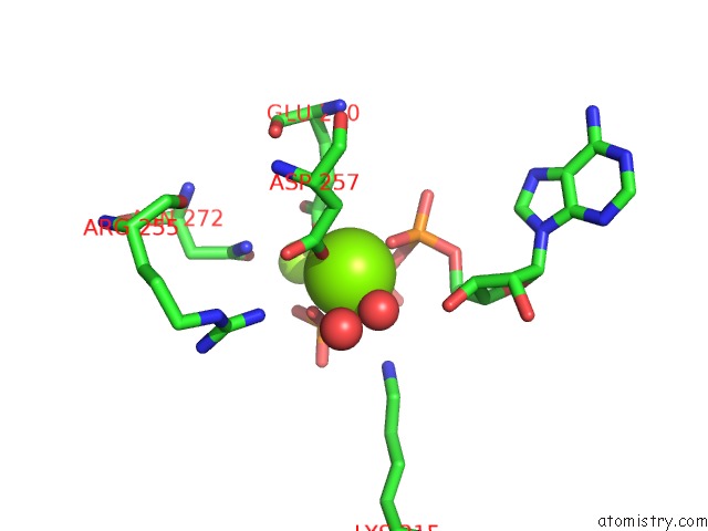
Mono view
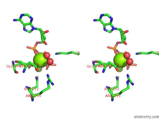
Stereo pair view
|
A full contact list of Magnesium with other atoms in the Mg binding
site number 2 of The X-Ray Crystal Structure of D-Alanyl-D-Alanine Ligase in Complex with Adp and D-Ala-D-Ala within 5.0Å range:
probe
|
atom
|
residue
|
distance (Å)
|
B
|
Occ
|
A:Mg1314
b:9.2
occ:1.00
|
O1A
|
A:ATP310
|
2.0
|
8.3
|
1.0
|
O2G
|
A:ATP310
|
2.0
|
8.6
|
1.0
|
OD2
|
A:ASP257
|
2.0
|
9.3
|
1.0
|
O
|
A:HOH2272
|
2.1
|
7.1
|
1.0
|
OE2
|
A:GLU270
|
2.1
|
6.5
|
1.0
|
O3B
|
A:ATP310
|
2.2
|
9.7
|
1.0
|
PG
|
A:ATP310
|
2.7
|
19.9
|
1.0
|
CD
|
A:GLU270
|
3.3
|
7.0
|
1.0
|
CG
|
A:ASP257
|
3.3
|
9.3
|
1.0
|
PA
|
A:ATP310
|
3.3
|
8.2
|
1.0
|
PB
|
A:ATP310
|
3.3
|
8.1
|
1.0
|
O3A
|
A:ATP310
|
3.6
|
8.5
|
1.0
|
O3G
|
A:ATP310
|
3.6
|
8.5
|
1.0
|
MG
|
A:MG1313
|
3.6
|
7.7
|
1.0
|
O1B
|
A:ATP310
|
3.8
|
7.2
|
1.0
|
CG
|
A:GLU270
|
3.8
|
7.4
|
1.0
|
NZ
|
A:LYS215
|
3.9
|
10.1
|
1.0
|
O1G
|
A:ATP310
|
4.0
|
9.2
|
1.0
|
ND2
|
A:ASN272
|
4.0
|
11.6
|
1.0
|
O
|
A:HOH2309
|
4.0
|
11.1
|
1.0
|
CB
|
A:ASP257
|
4.1
|
9.6
|
1.0
|
NH2
|
A:ARG255
|
4.1
|
10.3
|
1.0
|
OD1
|
A:ASP257
|
4.2
|
10.5
|
1.0
|
C5'
|
A:ATP310
|
4.2
|
10.2
|
1.0
|
O5'
|
A:ATP310
|
4.2
|
8.4
|
1.0
|
OE1
|
A:GLU270
|
4.3
|
7.5
|
1.0
|
O3'
|
A:ATP310
|
4.4
|
9.4
|
1.0
|
O2A
|
A:ATP310
|
4.4
|
9.1
|
1.0
|
O2B
|
A:ATP310
|
4.6
|
8.3
|
1.0
|
OD1
|
A:ASN272
|
4.7
|
9.2
|
1.0
|
CG
|
A:ASN272
|
4.8
|
10.0
|
1.0
|
C3'
|
A:ATP310
|
4.9
|
9.6
|
1.0
|
CE
|
A:LYS215
|
4.9
|
9.3
|
1.0
|
|
Magnesium binding site 3 out
of 7 in 4c5c
Go back to
Magnesium Binding Sites List in 4c5c
Magnesium binding site 3 out
of 7 in the The X-Ray Crystal Structure of D-Alanyl-D-Alanine Ligase in Complex with Adp and D-Ala-D-Ala
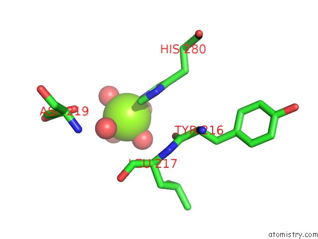
Mono view
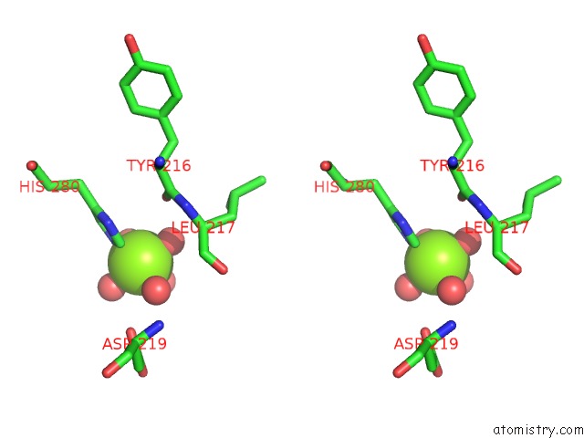
Stereo pair view
|
A full contact list of Magnesium with other atoms in the Mg binding
site number 3 of The X-Ray Crystal Structure of D-Alanyl-D-Alanine Ligase in Complex with Adp and D-Ala-D-Ala within 5.0Å range:
probe
|
atom
|
residue
|
distance (Å)
|
B
|
Occ
|
A:Mg1315
b:27.4
occ:1.00
|
O
|
A:HOH2273
|
1.9
|
26.5
|
1.0
|
O
|
A:HOH2322
|
2.0
|
20.4
|
1.0
|
O
|
A:HOH2323
|
2.0
|
23.5
|
1.0
|
NE2
|
A:HIS280
|
2.2
|
17.8
|
1.0
|
O
|
A:HOH2343
|
2.2
|
34.7
|
1.0
|
O
|
A:HOH2278
|
2.3
|
26.5
|
1.0
|
CE1
|
A:HIS280
|
3.0
|
15.1
|
1.0
|
CD2
|
A:HIS280
|
3.3
|
16.9
|
1.0
|
O
|
A:TYR216
|
4.1
|
11.9
|
1.0
|
ND1
|
A:HIS280
|
4.2
|
14.2
|
1.0
|
CG
|
A:HIS280
|
4.3
|
12.8
|
1.0
|
OD1
|
A:ASP219
|
4.5
|
32.0
|
1.0
|
CA
|
A:LEU217
|
4.9
|
13.8
|
1.0
|
|
Magnesium binding site 4 out
of 7 in 4c5c
Go back to
Magnesium Binding Sites List in 4c5c
Magnesium binding site 4 out
of 7 in the The X-Ray Crystal Structure of D-Alanyl-D-Alanine Ligase in Complex with Adp and D-Ala-D-Ala
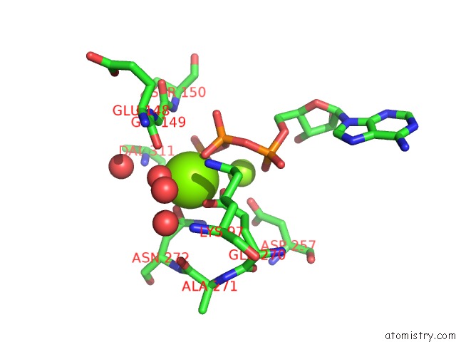
Mono view
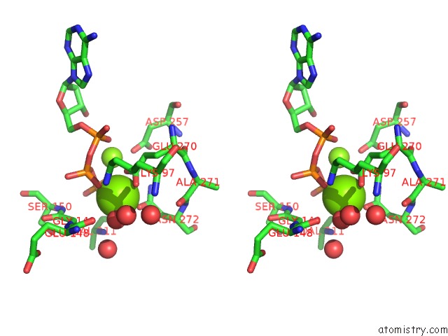
Stereo pair view
|
A full contact list of Magnesium with other atoms in the Mg binding
site number 4 of The X-Ray Crystal Structure of D-Alanyl-D-Alanine Ligase in Complex with Adp and D-Ala-D-Ala within 5.0Å range:
probe
|
atom
|
residue
|
distance (Å)
|
B
|
Occ
|
B:Mg1307
b:8.3
occ:1.00
|
O3G
|
B:ATP310
|
2.0
|
9.6
|
1.0
|
O1B
|
B:ATP310
|
2.0
|
8.6
|
1.0
|
O
|
B:HOH2165
|
2.1
|
4.8
|
1.0
|
OD1
|
B:ASN272
|
2.2
|
8.6
|
1.0
|
OE2
|
B:GLU270
|
2.2
|
6.7
|
1.0
|
OE1
|
B:GLU270
|
2.2
|
9.3
|
1.0
|
CD
|
B:GLU270
|
2.5
|
6.5
|
1.0
|
PG
|
B:ATP310
|
2.9
|
23.6
|
1.0
|
CG
|
B:ASN272
|
3.2
|
9.6
|
1.0
|
PB
|
B:ATP310
|
3.2
|
8.6
|
1.0
|
O3B
|
B:ATP310
|
3.3
|
10.0
|
1.0
|
O2G
|
B:ATP310
|
3.4
|
9.5
|
1.0
|
ND2
|
B:ASN272
|
3.6
|
11.7
|
1.0
|
MG
|
B:MG1308
|
3.6
|
9.3
|
1.0
|
O
|
B:HOH2105
|
3.6
|
2.0
|
1.0
|
CA
|
B:GLY149
|
4.0
|
9.3
|
1.0
|
CG
|
B:GLU270
|
4.0
|
8.3
|
1.0
|
NZ
|
B:LYS97
|
4.0
|
7.2
|
1.0
|
O1G
|
B:ATP310
|
4.1
|
10.4
|
1.0
|
O3A
|
B:ATP310
|
4.2
|
8.1
|
1.0
|
O
|
B:GLU148
|
4.2
|
8.8
|
1.0
|
O
|
B:HOH2018
|
4.2
|
10.3
|
1.0
|
N
|
B:DAL311
|
4.3
|
10.1
|
1.0
|
O2B
|
B:ATP310
|
4.3
|
8.3
|
1.0
|
CB
|
B:ASN272
|
4.5
|
9.5
|
1.0
|
O
|
B:HOH2106
|
4.6
|
7.6
|
1.0
|
O1A
|
B:ATP310
|
4.6
|
7.8
|
1.0
|
N
|
B:SER150
|
4.7
|
7.8
|
1.0
|
CE
|
B:LYS97
|
4.7
|
7.9
|
1.0
|
OD2
|
B:ASP257
|
4.8
|
9.2
|
1.0
|
C
|
B:GLY149
|
4.9
|
8.4
|
1.0
|
CB
|
B:GLU270
|
4.9
|
8.2
|
1.0
|
O
|
B:ALA271
|
5.0
|
10.8
|
1.0
|
C
|
B:GLU148
|
5.0
|
8.7
|
1.0
|
N
|
B:GLY149
|
5.0
|
9.1
|
1.0
|
|
Magnesium binding site 5 out
of 7 in 4c5c
Go back to
Magnesium Binding Sites List in 4c5c
Magnesium binding site 5 out
of 7 in the The X-Ray Crystal Structure of D-Alanyl-D-Alanine Ligase in Complex with Adp and D-Ala-D-Ala
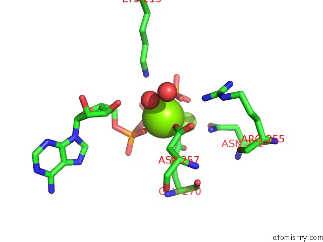
Mono view
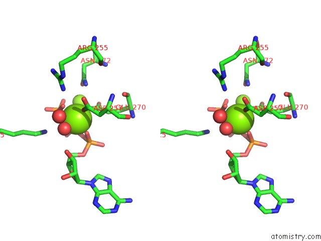
Stereo pair view
|
A full contact list of Magnesium with other atoms in the Mg binding
site number 5 of The X-Ray Crystal Structure of D-Alanyl-D-Alanine Ligase in Complex with Adp and D-Ala-D-Ala within 5.0Å range:
probe
|
atom
|
residue
|
distance (Å)
|
B
|
Occ
|
B:Mg1308
b:9.3
occ:1.00
|
O1A
|
B:ATP310
|
1.9
|
7.8
|
1.0
|
OE2
|
B:GLU270
|
2.1
|
6.7
|
1.0
|
OD2
|
B:ASP257
|
2.1
|
9.2
|
1.0
|
O3B
|
B:ATP310
|
2.1
|
10.0
|
1.0
|
O
|
B:HOH2236
|
2.1
|
7.9
|
1.0
|
O2G
|
B:ATP310
|
2.1
|
9.5
|
1.0
|
PG
|
B:ATP310
|
2.7
|
23.6
|
1.0
|
PA
|
B:ATP310
|
3.2
|
9.3
|
1.0
|
CD
|
B:GLU270
|
3.2
|
6.5
|
1.0
|
PB
|
B:ATP310
|
3.2
|
8.6
|
1.0
|
CG
|
B:ASP257
|
3.3
|
9.8
|
1.0
|
O3A
|
B:ATP310
|
3.5
|
8.1
|
1.0
|
MG
|
B:MG1307
|
3.6
|
8.3
|
1.0
|
O3G
|
B:ATP310
|
3.6
|
9.6
|
1.0
|
O1B
|
B:ATP310
|
3.7
|
8.6
|
1.0
|
CG
|
B:GLU270
|
3.8
|
8.3
|
1.0
|
NZ
|
B:LYS215
|
3.9
|
10.9
|
1.0
|
O1G
|
B:ATP310
|
3.9
|
10.4
|
1.0
|
ND2
|
B:ASN272
|
4.0
|
11.7
|
1.0
|
O
|
B:HOH2275
|
4.1
|
10.9
|
1.0
|
CB
|
B:ASP257
|
4.1
|
10.8
|
1.0
|
NH2
|
B:ARG255
|
4.1
|
11.1
|
1.0
|
C5'
|
B:ATP310
|
4.2
|
10.2
|
1.0
|
O5'
|
B:ATP310
|
4.2
|
8.3
|
1.0
|
OD1
|
B:ASP257
|
4.2
|
12.1
|
1.0
|
OE1
|
B:GLU270
|
4.2
|
9.3
|
1.0
|
O3'
|
B:ATP310
|
4.4
|
10.1
|
1.0
|
O2A
|
B:ATP310
|
4.4
|
10.1
|
1.0
|
O2B
|
B:ATP310
|
4.6
|
8.3
|
1.0
|
OD1
|
B:ASN272
|
4.6
|
8.6
|
1.0
|
CG
|
B:ASN272
|
4.8
|
9.6
|
1.0
|
CE
|
B:LYS215
|
4.9
|
10.1
|
1.0
|
C3'
|
B:ATP310
|
4.9
|
10.0
|
1.0
|
|
Magnesium binding site 6 out
of 7 in 4c5c
Go back to
Magnesium Binding Sites List in 4c5c
Magnesium binding site 6 out
of 7 in the The X-Ray Crystal Structure of D-Alanyl-D-Alanine Ligase in Complex with Adp and D-Ala-D-Ala
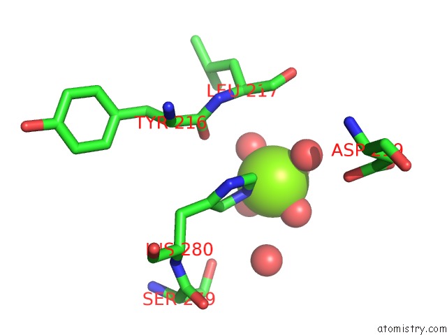
Mono view
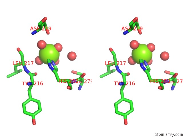
Stereo pair view
|
A full contact list of Magnesium with other atoms in the Mg binding
site number 6 of The X-Ray Crystal Structure of D-Alanyl-D-Alanine Ligase in Complex with Adp and D-Ala-D-Ala within 5.0Å range:
probe
|
atom
|
residue
|
distance (Å)
|
B
|
Occ
|
B:Mg1309
b:24.3
occ:1.00
|
O
|
B:HOH2241
|
1.8
|
22.9
|
1.0
|
O
|
B:HOH2286
|
1.9
|
17.0
|
1.0
|
O
|
B:HOH2238
|
2.0
|
23.6
|
1.0
|
NE2
|
B:HIS280
|
2.1
|
21.5
|
1.0
|
O
|
B:HOH2240
|
2.3
|
24.8
|
1.0
|
O
|
B:HOH2306
|
2.3
|
34.9
|
1.0
|
CE1
|
B:HIS280
|
3.0
|
20.8
|
1.0
|
CD2
|
B:HIS280
|
3.3
|
20.0
|
1.0
|
O
|
B:HOH2287
|
3.8
|
25.9
|
1.0
|
O
|
B:TYR216
|
4.1
|
13.9
|
1.0
|
ND1
|
B:HIS280
|
4.2
|
19.6
|
1.0
|
OD1
|
B:ASP219
|
4.3
|
32.9
|
1.0
|
CG
|
B:HIS280
|
4.3
|
15.5
|
1.0
|
CA
|
B:LEU217
|
4.8
|
17.3
|
1.0
|
OG
|
B:SER279
|
4.9
|
17.0
|
1.0
|
C
|
B:TYR216
|
5.0
|
12.8
|
1.0
|
|
Magnesium binding site 7 out
of 7 in 4c5c
Go back to
Magnesium Binding Sites List in 4c5c
Magnesium binding site 7 out
of 7 in the The X-Ray Crystal Structure of D-Alanyl-D-Alanine Ligase in Complex with Adp and D-Ala-D-Ala
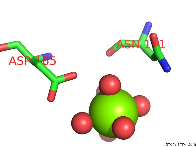
Mono view
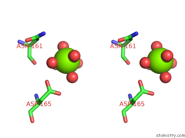
Stereo pair view
|
A full contact list of Magnesium with other atoms in the Mg binding
site number 7 of The X-Ray Crystal Structure of D-Alanyl-D-Alanine Ligase in Complex with Adp and D-Ala-D-Ala within 5.0Å range:
probe
|
atom
|
residue
|
distance (Å)
|
B
|
Occ
|
B:Mg1310
b:38.7
occ:1.00
|
O
|
B:HOH2308
|
2.0
|
26.7
|
1.0
|
O
|
B:HOH2307
|
2.0
|
20.0
|
1.0
|
O
|
B:HOH2182
|
2.1
|
20.3
|
1.0
|
O
|
B:HOH2187
|
2.3
|
15.2
|
1.0
|
O
|
B:HOH2181
|
2.5
|
25.6
|
1.0
|
O
|
B:HOH2186
|
2.6
|
32.6
|
1.0
|
OD2
|
B:ASP165
|
3.9
|
19.9
|
1.0
|
O
|
B:ASN161
|
4.4
|
21.1
|
1.0
|
OD1
|
B:ASP165
|
4.5
|
18.7
|
1.0
|
CG
|
B:ASP165
|
4.6
|
17.6
|
1.0
|
CB
|
B:ASN161
|
4.8
|
22.4
|
1.0
|
C
|
B:ASN161
|
5.0
|
19.5
|
1.0
|
ND2
|
B:ASN161
|
5.0
|
33.9
|
1.0
|
|
Reference:
S.Batson,
V.Majce,
A.J.Lloyd,
D.Rea,
C.W.G.Fishwick,
K.J.Simmons,
V.Fulop,
D.I.Roper.
D-Cycloserine Inhibition Occurs By A Novel Phosphoryl Intermediate: Prospects For Novel Antimicrobials Without Psychotic Side Effects. To Be Published.
Page generated: Thu Aug 15 16:42:41 2024
|