Magnesium »
PDB 4cgl-4cs4 »
4crz »
Magnesium in PDB 4crz: Direct Visualisation of Strain-Induced Protein Prost- Translational Modification
Enzymatic activity of Direct Visualisation of Strain-Induced Protein Prost- Translational Modification
All present enzymatic activity of Direct Visualisation of Strain-Induced Protein Prost- Translational Modification:
4.1.1.11;
4.1.1.11;
Protein crystallography data
The structure of Direct Visualisation of Strain-Induced Protein Prost- Translational Modification, PDB code: 4crz
was solved by
D.C.F.Monteiro,
V.Patel,
C.P.Bartlett,
T.D.Grant,
S.Nozaki,
J.A.Gowdy,
E.H.Snell,
H.Niki,
A.R.Pearson,
M.E.Webb,
with X-Ray Crystallography technique. A brief refinement statistics is given in the table below:
| Resolution Low / High (Å) | 29.52 / 1.70 |
| Space group | I 4 |
| Cell size a, b, c (Å), α, β, γ (°) | 86.280, 86.280, 80.880, 90.00, 90.00, 90.00 |
| R / Rfree (%) | 14.458 / 17.519 |
Magnesium Binding Sites:
The binding sites of Magnesium atom in the Direct Visualisation of Strain-Induced Protein Prost- Translational Modification
(pdb code 4crz). This binding sites where shown within
5.0 Angstroms radius around Magnesium atom.
In total only one binding site of Magnesium was determined in the Direct Visualisation of Strain-Induced Protein Prost- Translational Modification, PDB code: 4crz:
In total only one binding site of Magnesium was determined in the Direct Visualisation of Strain-Induced Protein Prost- Translational Modification, PDB code: 4crz:
Magnesium binding site 1 out of 1 in 4crz
Go back to
Magnesium binding site 1 out
of 1 in the Direct Visualisation of Strain-Induced Protein Prost- Translational Modification
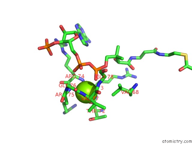
Mono view
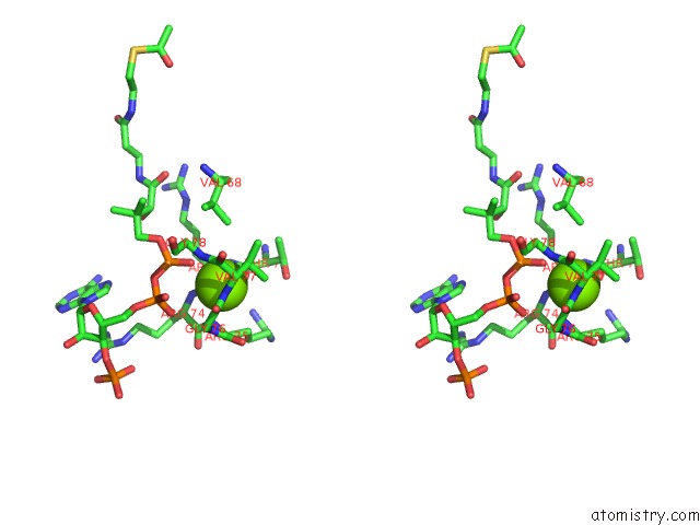
Stereo pair view
Mono view
Stereo pair view
A full contact list of Magnesium with other atoms in the Mg binding
site number 1 of Direct Visualisation of Strain-Induced Protein Prost- Translational Modification within 5.0Å range:
|
Reference:
D.C.F.Monteiro,
V.Patel,
C.P.Bartlett,
T.D.Grant,
S.Nozaki,
J.A.Gowdy,
E.H.Snell,
H.Niki,
A.R.Pearson,
M.E.Webb.
Direct Visualisation of Strain-Induced Protein Prost-Translational Modification To Be Published.
Page generated: Mon Aug 11 07:18:33 2025
Last articles
Mg in 4FNJMg in 4FPP
Mg in 4FP1
Mg in 4FO6
Mg in 4FO0
Mg in 4FME
Mg in 4FMM
Mg in 4FNI
Mg in 4FMO
Mg in 4FMD