Magnesium »
PDB 4d5e-4dhf »
4d6n »
Magnesium in PDB 4d6n: The Crystal Structure of I-Dmoi in Complex with Its Target Dna at 10 Days Incubation in 5MM Mg (State 7)
Protein crystallography data
The structure of The Crystal Structure of I-Dmoi in Complex with Its Target Dna at 10 Days Incubation in 5MM Mg (State 7), PDB code: 4d6n
was solved by
R.Molina,
S.Stella,
P.Redondo,
H.Gomez,
M.J.Marcaida,
M.Orozco,
J.Prieto,
G.Montoya,
with X-Ray Crystallography technique. A brief refinement statistics is given in the table below:
| Resolution Low / High (Å) | 46.598 / 2.35 |
| Space group | P 1 21 1 |
| Cell size a, b, c (Å), α, β, γ (°) | 106.311, 70.269, 107.169, 90.00, 119.59, 90.00 |
| R / Rfree (%) | 18.24 / 21.72 |
Magnesium Binding Sites:
The binding sites of Magnesium atom in the The Crystal Structure of I-Dmoi in Complex with Its Target Dna at 10 Days Incubation in 5MM Mg (State 7)
(pdb code 4d6n). This binding sites where shown within
5.0 Angstroms radius around Magnesium atom.
In total 6 binding sites of Magnesium where determined in the The Crystal Structure of I-Dmoi in Complex with Its Target Dna at 10 Days Incubation in 5MM Mg (State 7), PDB code: 4d6n:
Jump to Magnesium binding site number: 1; 2; 3; 4; 5; 6;
In total 6 binding sites of Magnesium where determined in the The Crystal Structure of I-Dmoi in Complex with Its Target Dna at 10 Days Incubation in 5MM Mg (State 7), PDB code: 4d6n:
Jump to Magnesium binding site number: 1; 2; 3; 4; 5; 6;
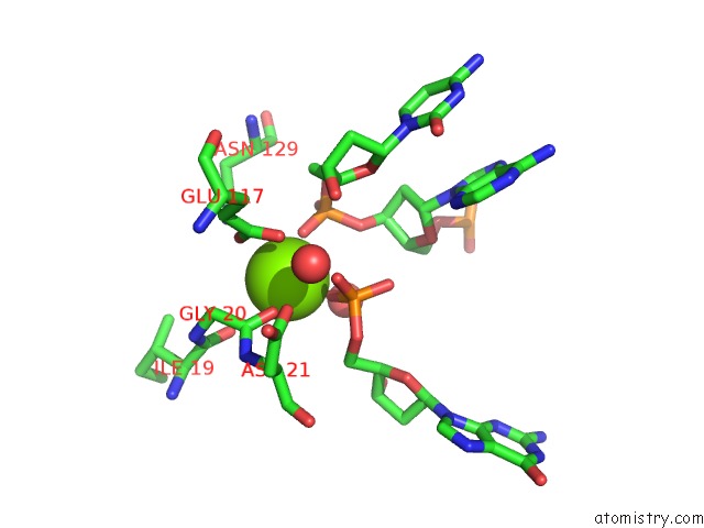
Magnesium binding site 1 out of 6 in 4d6n
Go back to
Magnesium binding site 1 out
of 6 in the The Crystal Structure of I-Dmoi in Complex with Its Target Dna at 10 Days Incubation in 5MM Mg (State 7)

Mono view
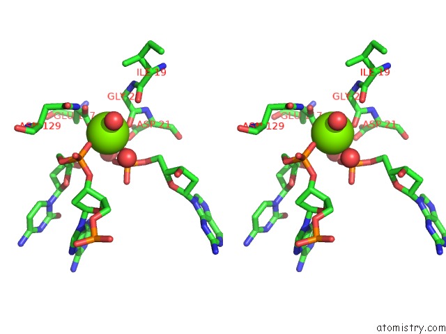
Stereo pair view
Mono view
Stereo pair view
A full contact list of Magnesium with other atoms in the Mg binding
site number 1 of The Crystal Structure of I-Dmoi in Complex with Its Target Dna at 10 Days Incubation in 5MM Mg (State 7) within 5.0Å range:
|
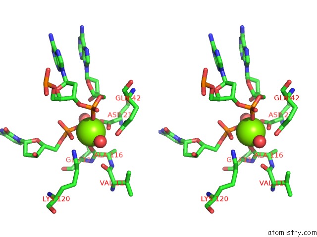
Magnesium binding site 2 out of 6 in 4d6n
Go back to
Magnesium binding site 2 out
of 6 in the The Crystal Structure of I-Dmoi in Complex with Its Target Dna at 10 Days Incubation in 5MM Mg (State 7)

Mono view

Stereo pair view
Mono view
Stereo pair view
A full contact list of Magnesium with other atoms in the Mg binding
site number 2 of The Crystal Structure of I-Dmoi in Complex with Its Target Dna at 10 Days Incubation in 5MM Mg (State 7) within 5.0Å range:
|
Magnesium binding site 3 out of 6 in 4d6n
Go back to
Magnesium binding site 3 out
of 6 in the The Crystal Structure of I-Dmoi in Complex with Its Target Dna at 10 Days Incubation in 5MM Mg (State 7)

Mono view
Stereo pair view
Mono view
Stereo pair view
A full contact list of Magnesium with other atoms in the Mg binding
site number 3 of The Crystal Structure of I-Dmoi in Complex with Its Target Dna at 10 Days Incubation in 5MM Mg (State 7) within 5.0Å range:
|
Magnesium binding site 4 out of 6 in 4d6n
Go back to
Magnesium binding site 4 out
of 6 in the The Crystal Structure of I-Dmoi in Complex with Its Target Dna at 10 Days Incubation in 5MM Mg (State 7)

Mono view

Stereo pair view
Mono view
Stereo pair view
A full contact list of Magnesium with other atoms in the Mg binding
site number 4 of The Crystal Structure of I-Dmoi in Complex with Its Target Dna at 10 Days Incubation in 5MM Mg (State 7) within 5.0Å range:
|
Magnesium binding site 5 out of 6 in 4d6n
Go back to
Magnesium binding site 5 out
of 6 in the The Crystal Structure of I-Dmoi in Complex with Its Target Dna at 10 Days Incubation in 5MM Mg (State 7)
Mono view

Stereo pair view
Mono view
Stereo pair view
A full contact list of Magnesium with other atoms in the Mg binding
site number 5 of The Crystal Structure of I-Dmoi in Complex with Its Target Dna at 10 Days Incubation in 5MM Mg (State 7) within 5.0Å range:
|
Magnesium binding site 6 out of 6 in 4d6n
Go back to
Magnesium binding site 6 out
of 6 in the The Crystal Structure of I-Dmoi in Complex with Its Target Dna at 10 Days Incubation in 5MM Mg (State 7)

Mono view

Stereo pair view
Mono view
Stereo pair view
A full contact list of Magnesium with other atoms in the Mg binding
site number 6 of The Crystal Structure of I-Dmoi in Complex with Its Target Dna at 10 Days Incubation in 5MM Mg (State 7) within 5.0Å range:
|
Reference:
R.Molina,
S.Stella,
P.Redondo,
H.Gomez,
M.J.Marcaida,
M.Orozco,
J.Prieto,
G.Montoya.
Visualizing Phosphodiester-Bond Hydrolysis By An Endonuclease. Nat.Struct.Mol.Biol. 2014.
ISSN: ESSN 1545-9985
PubMed: 25486305
DOI: 10.1038/NSMB.2932
Page generated: Mon Aug 11 07:24:38 2025
ISSN: ESSN 1545-9985
PubMed: 25486305
DOI: 10.1038/NSMB.2932
Last articles
Mg in 4FE3Mg in 4FE2
Mg in 4FCE
Mg in 4FCD
Mg in 4FCB
Mg in 4FAW
Mg in 4FAX
Mg in 4FAR
Mg in 4FAU
Mg in 4F86