Magnesium »
PDB 4d5e-4dhf »
4da1 »
Magnesium in PDB 4da1: Crystal Structure of Branched-Chain Alpha-Ketoacid Dehydrogenase Phosphatase with Mg (II) Ions at the Active Site
Enzymatic activity of Crystal Structure of Branched-Chain Alpha-Ketoacid Dehydrogenase Phosphatase with Mg (II) Ions at the Active Site
All present enzymatic activity of Crystal Structure of Branched-Chain Alpha-Ketoacid Dehydrogenase Phosphatase with Mg (II) Ions at the Active Site:
3.1.3.16;
3.1.3.16;
Protein crystallography data
The structure of Crystal Structure of Branched-Chain Alpha-Ketoacid Dehydrogenase Phosphatase with Mg (II) Ions at the Active Site, PDB code: 4da1
was solved by
C.A.Brautigam,
J.L.Chuang,
D.T.Chuang,
with X-Ray Crystallography technique. A brief refinement statistics is given in the table below:
| Resolution Low / High (Å) | 43.94 / 2.38 |
| Space group | P 31 2 1 |
| Cell size a, b, c (Å), α, β, γ (°) | 125.276, 125.276, 61.660, 90.00, 90.00, 120.00 |
| R / Rfree (%) | 17.7 / 20.5 |
Magnesium Binding Sites:
The binding sites of Magnesium atom in the Crystal Structure of Branched-Chain Alpha-Ketoacid Dehydrogenase Phosphatase with Mg (II) Ions at the Active Site
(pdb code 4da1). This binding sites where shown within
5.0 Angstroms radius around Magnesium atom.
In total 3 binding sites of Magnesium where determined in the Crystal Structure of Branched-Chain Alpha-Ketoacid Dehydrogenase Phosphatase with Mg (II) Ions at the Active Site, PDB code: 4da1:
Jump to Magnesium binding site number: 1; 2; 3;
In total 3 binding sites of Magnesium where determined in the Crystal Structure of Branched-Chain Alpha-Ketoacid Dehydrogenase Phosphatase with Mg (II) Ions at the Active Site, PDB code: 4da1:
Jump to Magnesium binding site number: 1; 2; 3;
Magnesium binding site 1 out of 3 in 4da1
Go back to
Magnesium binding site 1 out
of 3 in the Crystal Structure of Branched-Chain Alpha-Ketoacid Dehydrogenase Phosphatase with Mg (II) Ions at the Active Site

Mono view
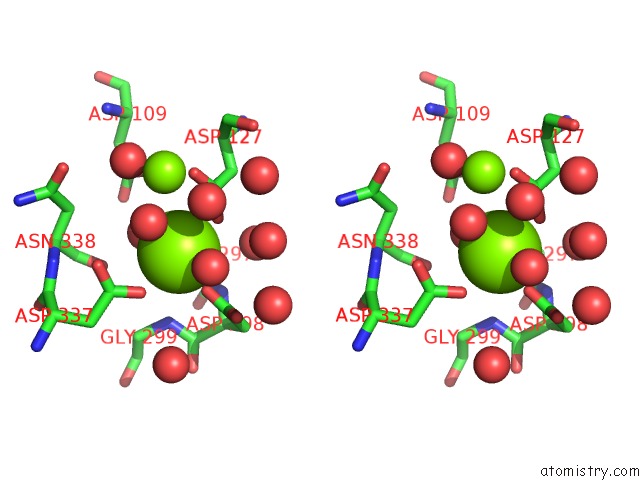
Stereo pair view
Mono view
Stereo pair view
A full contact list of Magnesium with other atoms in the Mg binding
site number 1 of Crystal Structure of Branched-Chain Alpha-Ketoacid Dehydrogenase Phosphatase with Mg (II) Ions at the Active Site within 5.0Å range:
|
Magnesium binding site 2 out of 3 in 4da1
Go back to
Magnesium binding site 2 out
of 3 in the Crystal Structure of Branched-Chain Alpha-Ketoacid Dehydrogenase Phosphatase with Mg (II) Ions at the Active Site

Mono view

Stereo pair view
Mono view
Stereo pair view
A full contact list of Magnesium with other atoms in the Mg binding
site number 2 of Crystal Structure of Branched-Chain Alpha-Ketoacid Dehydrogenase Phosphatase with Mg (II) Ions at the Active Site within 5.0Å range:
|
Magnesium binding site 3 out of 3 in 4da1
Go back to
Magnesium binding site 3 out
of 3 in the Crystal Structure of Branched-Chain Alpha-Ketoacid Dehydrogenase Phosphatase with Mg (II) Ions at the Active Site

Mono view

Stereo pair view
Mono view
Stereo pair view
A full contact list of Magnesium with other atoms in the Mg binding
site number 3 of Crystal Structure of Branched-Chain Alpha-Ketoacid Dehydrogenase Phosphatase with Mg (II) Ions at the Active Site within 5.0Å range:
|
Reference:
R.M.Wynn,
J.Li,
C.A.Brautigam,
J.L.Chuang,
D.T.Chuang.
Structural and Biochemical Characterization of Human Mitochondrial Branched-Chain Alpha-Ketoacid Dehydrogenase Phosphatase. J.Biol.Chem. V. 287 9178 2012.
ISSN: ISSN 0021-9258
PubMed: 22291014
DOI: 10.1074/JBC.M111.314963
Page generated: Mon Aug 11 07:26:00 2025
ISSN: ISSN 0021-9258
PubMed: 22291014
DOI: 10.1074/JBC.M111.314963
Last articles
Mg in 4FAUMg in 4F86
Mg in 4F9A
Mg in 4F99
Mg in 4F93
Mg in 4F8B
Mg in 4F8E
Mg in 4F71
Mg in 4F72
Mg in 4F6X