Magnesium »
PDB 4d5e-4dhf »
4dbf »
Magnesium in PDB 4dbf: Crystal Structures of CG1458
Protein crystallography data
The structure of Crystal Structures of CG1458, PDB code: 4dbf
was solved by
T.T.Ran,
D.Q.Xu,
W.W.Wang,
Y.Y.Gao,
M.T.Wang,
with X-Ray Crystallography technique. A brief refinement statistics is given in the table below:
| Resolution Low / High (Å) | 20.00 / 1.90 |
| Space group | P 43 21 2 |
| Cell size a, b, c (Å), α, β, γ (°) | 124.120, 124.120, 73.640, 90.00, 90.00, 90.00 |
| R / Rfree (%) | 19.4 / 24.4 |
Magnesium Binding Sites:
The binding sites of Magnesium atom in the Crystal Structures of CG1458
(pdb code 4dbf). This binding sites where shown within
5.0 Angstroms radius around Magnesium atom.
In total 3 binding sites of Magnesium where determined in the Crystal Structures of CG1458, PDB code: 4dbf:
Jump to Magnesium binding site number: 1; 2; 3;
In total 3 binding sites of Magnesium where determined in the Crystal Structures of CG1458, PDB code: 4dbf:
Jump to Magnesium binding site number: 1; 2; 3;
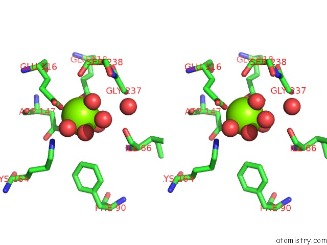
Magnesium binding site 1 out of 3 in 4dbf
Go back to
Magnesium binding site 1 out
of 3 in the Crystal Structures of CG1458

Mono view
Stereo pair view
Mono view
Stereo pair view
A full contact list of Magnesium with other atoms in the Mg binding
site number 1 of Crystal Structures of CG1458 within 5.0Å range:
|
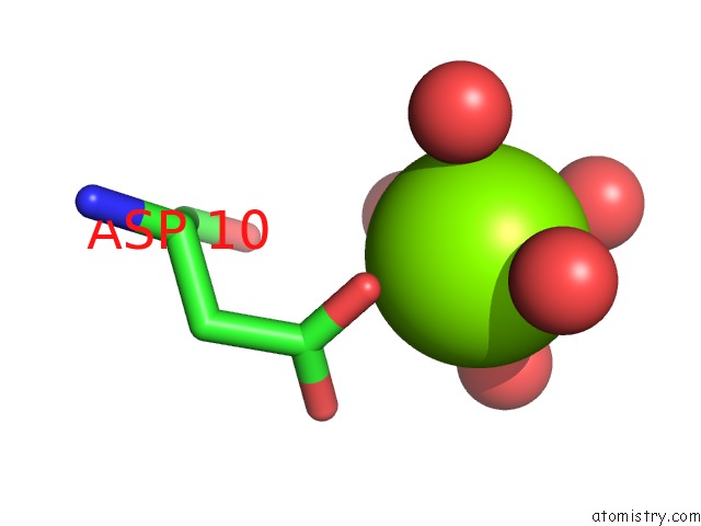
Magnesium binding site 2 out of 3 in 4dbf
Go back to
Magnesium binding site 2 out
of 3 in the Crystal Structures of CG1458
Mono view

Stereo pair view
Mono view
Stereo pair view
A full contact list of Magnesium with other atoms in the Mg binding
site number 2 of Crystal Structures of CG1458 within 5.0Å range:
|
Magnesium binding site 3 out of 3 in 4dbf
Go back to
Magnesium binding site 3 out
of 3 in the Crystal Structures of CG1458

Mono view

Stereo pair view
Mono view
Stereo pair view
A full contact list of Magnesium with other atoms in the Mg binding
site number 3 of Crystal Structures of CG1458 within 5.0Å range:
|
Reference:
T.Ran,
Y.Gao,
M.Marsh,
W.Zhu,
M.Wang,
X.Mao,
L.Xu,
D.Xu,
W.Wang.
Crystal Structures of CG1458 Reveal A Catalytic Lid Domain and A Common Catalytic Mechanism For Fah Family. Biochem.J. V. 449 51 2013.
ISSN: ISSN 0264-6021
PubMed: 23046410
DOI: 10.1042/BJ20120913
Page generated: Mon Aug 11 07:26:16 2025
ISSN: ISSN 0264-6021
PubMed: 23046410
DOI: 10.1042/BJ20120913
Last articles
Mg in 4FAUMg in 4F86
Mg in 4F9A
Mg in 4F99
Mg in 4F93
Mg in 4F8B
Mg in 4F8E
Mg in 4F71
Mg in 4F72
Mg in 4F6X