Magnesium »
PDB 4dhf-4do9 »
4dn5 »
Magnesium in PDB 4dn5: Crystal Structure of Nf-Kb-Inducing Kinase (Nik)
Enzymatic activity of Crystal Structure of Nf-Kb-Inducing Kinase (Nik)
All present enzymatic activity of Crystal Structure of Nf-Kb-Inducing Kinase (Nik):
2.7.11.25;
2.7.11.25;
Protein crystallography data
The structure of Crystal Structure of Nf-Kb-Inducing Kinase (Nik), PDB code: 4dn5
was solved by
X.Min,
J.Liu,
A.Sudom,
N.P.Walker,
Z.Wang,
with X-Ray Crystallography technique. A brief refinement statistics is given in the table below:
| Resolution Low / High (Å) | 30.00 / 2.50 |
| Space group | P 41 |
| Cell size a, b, c (Å), α, β, γ (°) | 85.060, 85.060, 115.400, 90.00, 90.00, 90.00 |
| R / Rfree (%) | 18.2 / 22.2 |
Magnesium Binding Sites:
The binding sites of Magnesium atom in the Crystal Structure of Nf-Kb-Inducing Kinase (Nik)
(pdb code 4dn5). This binding sites where shown within
5.0 Angstroms radius around Magnesium atom.
In total 2 binding sites of Magnesium where determined in the Crystal Structure of Nf-Kb-Inducing Kinase (Nik), PDB code: 4dn5:
Jump to Magnesium binding site number: 1; 2;
In total 2 binding sites of Magnesium where determined in the Crystal Structure of Nf-Kb-Inducing Kinase (Nik), PDB code: 4dn5:
Jump to Magnesium binding site number: 1; 2;
Magnesium binding site 1 out of 2 in 4dn5
Go back to
Magnesium binding site 1 out
of 2 in the Crystal Structure of Nf-Kb-Inducing Kinase (Nik)
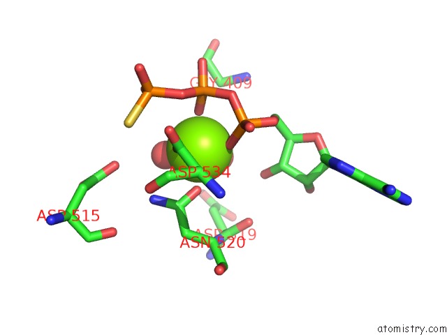
Mono view
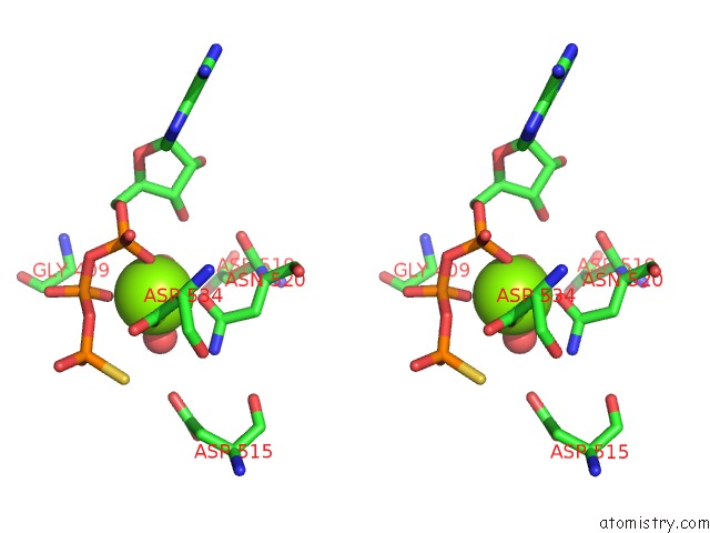
Stereo pair view
Mono view
Stereo pair view
A full contact list of Magnesium with other atoms in the Mg binding
site number 1 of Crystal Structure of Nf-Kb-Inducing Kinase (Nik) within 5.0Å range:
|
Magnesium binding site 2 out of 2 in 4dn5
Go back to
Magnesium binding site 2 out
of 2 in the Crystal Structure of Nf-Kb-Inducing Kinase (Nik)
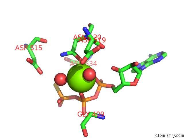
Mono view

Stereo pair view
Mono view
Stereo pair view
A full contact list of Magnesium with other atoms in the Mg binding
site number 2 of Crystal Structure of Nf-Kb-Inducing Kinase (Nik) within 5.0Å range:
|
Reference:
J.Liu,
A.Sudom,
X.Min,
Z.Cao,
X.Gao,
M.Ayres,
F.Lee,
P.Cao,
S.Johnstone,
O.Plotnikova,
N.Walker,
G.Chen,
Z.Wang.
Structure of Nuclear Factor Kappa B-Inducing Kinase Domain Reveals A Constitutively Active Conformation J.Biol.Chem. V. 287 27326 2012.
ISSN: ISSN 0021-9258
PubMed: 22718757
DOI: 10.1074/JBC.M112.366658
Page generated: Thu Aug 15 17:16:35 2024
ISSN: ISSN 0021-9258
PubMed: 22718757
DOI: 10.1074/JBC.M112.366658
Last articles
Zn in 9J0NZn in 9J0O
Zn in 9J0P
Zn in 9FJX
Zn in 9EKB
Zn in 9C0F
Zn in 9CAH
Zn in 9CH0
Zn in 9CH3
Zn in 9CH1