Magnesium »
PDB 4e7z-4egh »
4ee0 »
Magnesium in PDB 4ee0: Crystal Structure of Hh-Pgds with Water Displacing Inhibitor
Enzymatic activity of Crystal Structure of Hh-Pgds with Water Displacing Inhibitor
All present enzymatic activity of Crystal Structure of Hh-Pgds with Water Displacing Inhibitor:
2.5.1.18; 5.3.99.2;
2.5.1.18; 5.3.99.2;
Protein crystallography data
The structure of Crystal Structure of Hh-Pgds with Water Displacing Inhibitor, PDB code: 4ee0
was solved by
J.E.Day,
A.Thorarensen,
J.I.Trujillo,
with X-Ray Crystallography technique. A brief refinement statistics is given in the table below:
| Resolution Low / High (Å) | 19.73 / 1.75 |
| Space group | P 1 21 1 |
| Cell size a, b, c (Å), α, β, γ (°) | 48.612, 77.876, 52.560, 90.00, 91.39, 90.00 |
| R / Rfree (%) | 19.6 / 22.3 |
Magnesium Binding Sites:
The binding sites of Magnesium atom in the Crystal Structure of Hh-Pgds with Water Displacing Inhibitor
(pdb code 4ee0). This binding sites where shown within
5.0 Angstroms radius around Magnesium atom.
In total only one binding site of Magnesium was determined in the Crystal Structure of Hh-Pgds with Water Displacing Inhibitor, PDB code: 4ee0:
In total only one binding site of Magnesium was determined in the Crystal Structure of Hh-Pgds with Water Displacing Inhibitor, PDB code: 4ee0:
Magnesium binding site 1 out of 1 in 4ee0
Go back to
Magnesium binding site 1 out
of 1 in the Crystal Structure of Hh-Pgds with Water Displacing Inhibitor
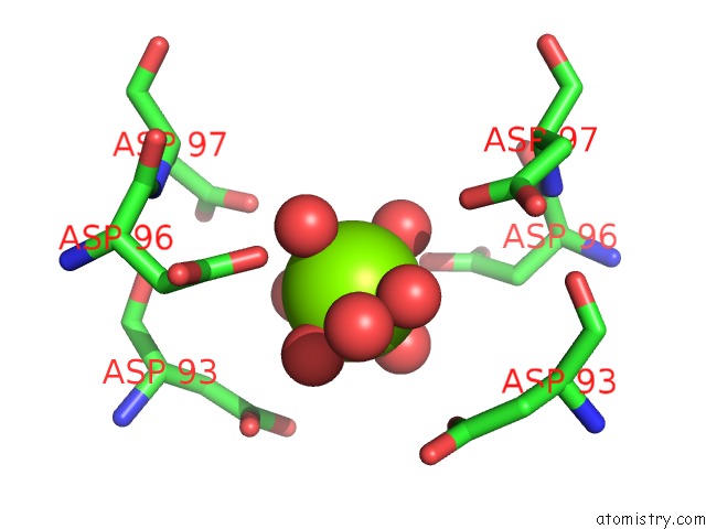
Mono view
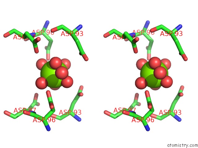
Stereo pair view
Mono view
Stereo pair view
A full contact list of Magnesium with other atoms in the Mg binding
site number 1 of Crystal Structure of Hh-Pgds with Water Displacing Inhibitor within 5.0Å range:
|
Reference:
J.I.Trujillo,
J.R.Kiefer,
W.Huang,
J.E.Day,
J.Moon,
G.M.Jerome,
C.P.Bono,
C.M.Kornmeier,
M.L.Williams,
C.Kuhn,
G.R.Rennie,
T.A.Wynn,
C.P.Carron,
A.Thorarensen.
Investigation of the Binding Pocket of Human Hematopoietic Prostaglandin (Pg) D2 Synthase (Hh-Pgds): A Tale of Two Waters. Bioorg.Med.Chem.Lett. V. 22 3795 2012.
ISSN: ISSN 0960-894X
PubMed: 22546671
DOI: 10.1016/J.BMCL.2012.04.004
Page generated: Fri Aug 16 14:28:39 2024
ISSN: ISSN 0960-894X
PubMed: 22546671
DOI: 10.1016/J.BMCL.2012.04.004
Last articles
Zn in 9JYWZn in 9IR4
Zn in 9IR3
Zn in 9GMX
Zn in 9GMW
Zn in 9JEJ
Zn in 9ERF
Zn in 9ERE
Zn in 9EGV
Zn in 9EGW