Magnesium in PDB 4fk9: High Resolution Structure of the Catalytic Domain of Mannanase SACTE_2347 From Streptomyces Sp. Sirexaa-E
Protein crystallography data
The structure of High Resolution Structure of the Catalytic Domain of Mannanase SACTE_2347 From Streptomyces Sp. Sirexaa-E, PDB code: 4fk9
was solved by
J.F.Acheson,
T.E.Takasuka,
B.G.Fox,
with X-Ray Crystallography technique. A brief refinement statistics is given in the table below:
Resolution Low / High (Å)
|
36.77 /
1.06
|
Space group
|
P 21 21 2
|
Cell size a, b, c (Å), α, β, γ (°)
|
62.805,
102.360,
45.346,
90.00,
90.00,
90.00
|
R / Rfree (%)
|
11.9 /
13.2
|
Other elements in 4fk9:
The structure of High Resolution Structure of the Catalytic Domain of Mannanase SACTE_2347 From Streptomyces Sp. Sirexaa-E also contains other interesting chemical elements:
Magnesium Binding Sites:
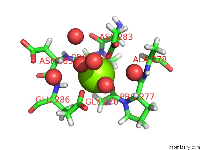
The binding sites of Magnesium atom in the High Resolution Structure of the Catalytic Domain of Mannanase SACTE_2347 From Streptomyces Sp. Sirexaa-E
(pdb code 4fk9). This binding sites where shown within
5.0 Angstroms radius around Magnesium atom.
In total 5 binding sites of Magnesium where determined in the
High Resolution Structure of the Catalytic Domain of Mannanase SACTE_2347 From Streptomyces Sp. Sirexaa-E, PDB code: 4fk9:
Jump to Magnesium binding site number:
1;
2;
3;
4;
5;
Magnesium binding site 1 out
of 5 in 4fk9
Go back to
Magnesium Binding Sites List in 4fk9
Magnesium binding site 1 out
of 5 in the High Resolution Structure of the Catalytic Domain of Mannanase SACTE_2347 From Streptomyces Sp. Sirexaa-E
Mono view
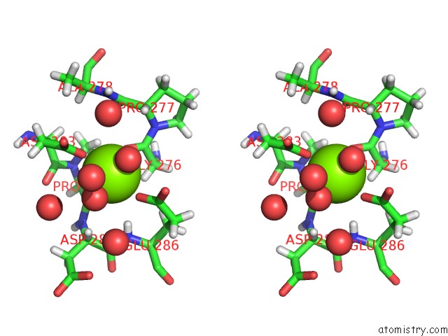
Stereo pair view
|
A full contact list of Magnesium with other atoms in the Mg binding
site number 1 of High Resolution Structure of the Catalytic Domain of Mannanase SACTE_2347 From Streptomyces Sp. Sirexaa-E within 5.0Å range:
probe
|
atom
|
residue
|
distance (Å)
|
B
|
Occ
|
A:Mg401
b:12.4
occ:1.00
|
OD1
|
A:ASP283
|
2.0
|
15.3
|
1.0
|
O
|
A:GLY276
|
2.1
|
13.0
|
1.0
|
O
|
A:HOH619
|
2.1
|
16.0
|
1.0
|
O
|
A:PRO284
|
2.1
|
11.8
|
1.0
|
O
|
A:HOH579
|
2.1
|
14.4
|
1.0
|
OE1
|
A:GLU286
|
2.1
|
12.9
|
1.0
|
C
|
A:GLY276
|
3.1
|
12.5
|
1.0
|
CG
|
A:ASP283
|
3.2
|
16.1
|
1.0
|
CD
|
A:GLU286
|
3.2
|
13.4
|
1.0
|
HA2
|
A:GLY276
|
3.2
|
14.7
|
1.0
|
C
|
A:PRO284
|
3.3
|
11.6
|
1.0
|
H
|
A:GLU286
|
3.4
|
13.2
|
1.0
|
OE2
|
A:GLU286
|
3.5
|
15.9
|
1.0
|
HD2
|
A:PRO284
|
3.6
|
15.7
|
1.0
|
CA
|
A:GLY276
|
3.7
|
12.2
|
1.0
|
OD2
|
A:ASP283
|
3.7
|
17.9
|
1.0
|
HA
|
A:ASP285
|
3.7
|
13.9
|
1.0
|
O
|
A:HOH913
|
3.9
|
36.1
|
1.0
|
N
|
A:PRO284
|
3.9
|
13.3
|
1.0
|
C
|
A:ASP283
|
4.0
|
14.6
|
1.0
|
O
|
A:HOH865
|
4.0
|
28.5
|
1.0
|
HA3
|
A:GLY276
|
4.1
|
14.7
|
1.0
|
O
|
A:HOH593
|
4.1
|
20.9
|
1.0
|
HB2
|
A:PRO284
|
4.1
|
14.4
|
1.0
|
HA
|
A:PRO277
|
4.1
|
17.4
|
1.0
|
CA
|
A:PRO284
|
4.1
|
12.2
|
1.0
|
N
|
A:PRO277
|
4.2
|
13.2
|
1.0
|
N
|
A:GLU286
|
4.2
|
11.0
|
1.0
|
O
|
A:HOH582
|
4.2
|
13.8
|
1.0
|
N
|
A:ASP285
|
4.3
|
11.5
|
1.0
|
CD
|
A:PRO284
|
4.3
|
13.1
|
1.0
|
O
|
A:ASP283
|
4.3
|
15.0
|
1.0
|
CB
|
A:ASP283
|
4.4
|
16.1
|
1.0
|
HA
|
A:ASP283
|
4.4
|
19.3
|
1.0
|
HB3
|
A:GLU286
|
4.4
|
13.8
|
1.0
|
CA
|
A:ASP285
|
4.4
|
11.6
|
1.0
|
CA
|
A:PRO277
|
4.5
|
14.5
|
1.0
|
CA
|
A:ASP283
|
4.5
|
16.1
|
1.0
|
CG
|
A:GLU286
|
4.5
|
12.5
|
1.0
|
CB
|
A:PRO284
|
4.6
|
12.0
|
1.0
|
C
|
A:PRO277
|
4.6
|
14.9
|
1.0
|
HB3
|
A:ASP283
|
4.7
|
19.3
|
1.0
|
H
|
A:ALA278
|
4.8
|
19.8
|
1.0
|
N
|
A:ALA278
|
4.8
|
16.5
|
1.0
|
C
|
A:ASP285
|
4.9
|
11.0
|
1.0
|
CB
|
A:GLU286
|
4.9
|
11.5
|
1.0
|
N
|
A:GLY276
|
4.9
|
11.7
|
1.0
|
HG2
|
A:GLU286
|
5.0
|
14.9
|
1.0
|
|
Magnesium binding site 2 out
of 5 in 4fk9
Go back to
Magnesium Binding Sites List in 4fk9
Magnesium binding site 2 out
of 5 in the High Resolution Structure of the Catalytic Domain of Mannanase SACTE_2347 From Streptomyces Sp. Sirexaa-E
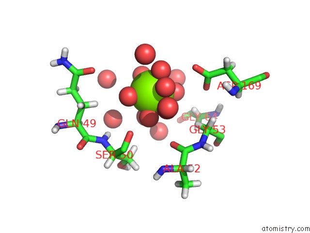
Mono view
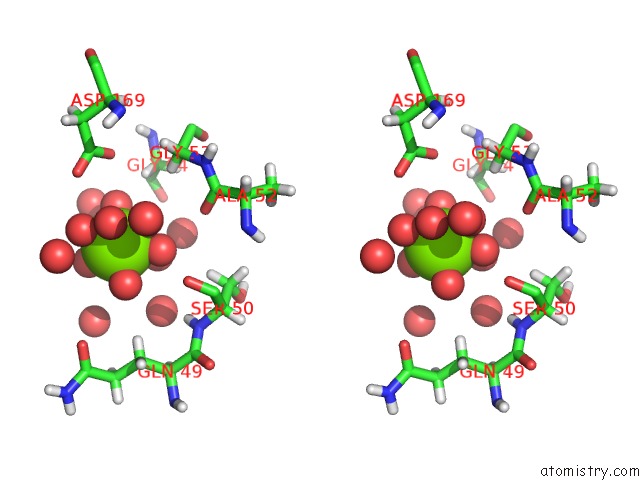
Stereo pair view
|
A full contact list of Magnesium with other atoms in the Mg binding
site number 2 of High Resolution Structure of the Catalytic Domain of Mannanase SACTE_2347 From Streptomyces Sp. Sirexaa-E within 5.0Å range:
probe
|
atom
|
residue
|
distance (Å)
|
B
|
Occ
|
A:Mg402
b:11.2
occ:0.79
|
O
|
A:HOH707
|
2.0
|
20.6
|
1.0
|
O
|
A:HOH606
|
2.0
|
20.0
|
1.0
|
O
|
A:HOH657
|
2.0
|
24.7
|
1.0
|
O
|
A:HOH996
|
2.1
|
23.2
|
1.0
|
O
|
A:HOH986
|
2.1
|
18.7
|
1.0
|
O
|
A:HOH572
|
2.1
|
13.8
|
1.0
|
O
|
A:HOH852
|
2.2
|
29.4
|
1.0
|
O
|
A:HOH821
|
2.3
|
23.0
|
1.0
|
O
|
A:HOH987
|
2.4
|
27.2
|
1.0
|
OD2
|
A:ASP169
|
4.1
|
9.8
|
1.0
|
O
|
A:HOH655
|
4.1
|
20.8
|
1.0
|
O
|
A:SER50
|
4.2
|
11.9
|
1.0
|
OD1
|
A:ASP169
|
4.2
|
8.0
|
1.0
|
O
|
A:HOH701
|
4.2
|
20.0
|
1.0
|
O
|
A:HOH584
|
4.3
|
17.8
|
1.0
|
HA2
|
A:GLY53
|
4.3
|
7.6
|
1.0
|
O
|
A:HOH1003
|
4.3
|
38.0
|
1.0
|
O
|
A:ALA52
|
4.4
|
8.8
|
1.0
|
O
|
A:HOH717
|
4.4
|
31.3
|
1.0
|
HB3
|
A:GLN49
|
4.5
|
21.0
|
1.0
|
HB2
|
A:SER50
|
4.6
|
19.9
|
1.0
|
O
|
A:HOH704
|
4.6
|
23.7
|
1.0
|
CG
|
A:ASP169
|
4.6
|
7.7
|
1.0
|
HA3
|
A:GLY64
|
4.7
|
12.2
|
1.0
|
H
|
A:SER50
|
4.7
|
16.6
|
1.0
|
OE1
|
A:GLN49
|
5.0
|
25.4
|
1.0
|
|
Magnesium binding site 3 out
of 5 in 4fk9
Go back to
Magnesium Binding Sites List in 4fk9
Magnesium binding site 3 out
of 5 in the High Resolution Structure of the Catalytic Domain of Mannanase SACTE_2347 From Streptomyces Sp. Sirexaa-E
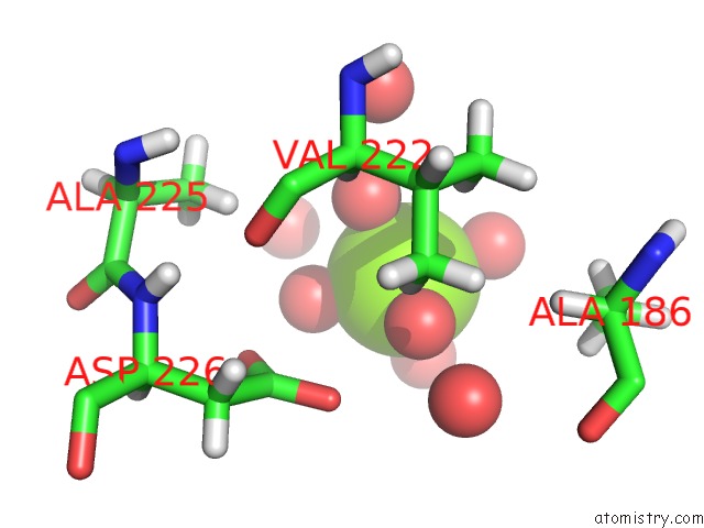
Mono view
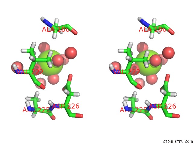
Stereo pair view
|
A full contact list of Magnesium with other atoms in the Mg binding
site number 3 of High Resolution Structure of the Catalytic Domain of Mannanase SACTE_2347 From Streptomyces Sp. Sirexaa-E within 5.0Å range:
probe
|
atom
|
residue
|
distance (Å)
|
B
|
Occ
|
A:Mg403
b:10.8
occ:0.34
|
O
|
A:HOH700
|
2.0
|
20.5
|
1.0
|
O
|
A:HOH706
|
2.1
|
23.3
|
1.0
|
O
|
A:HOH848
|
2.1
|
24.1
|
1.0
|
O
|
A:HOH847
|
2.1
|
31.0
|
1.0
|
O
|
A:HOH841
|
2.1
|
23.1
|
1.0
|
O
|
A:HOH849
|
2.2
|
29.6
|
1.0
|
O
|
A:HOH936
|
3.2
|
27.7
|
1.0
|
O
|
A:HOH711
|
3.8
|
22.5
|
1.0
|
HG22
|
A:VAL222
|
4.1
|
10.8
|
1.0
|
OD1
|
A:ASP226
|
4.2
|
11.6
|
1.0
|
OD2
|
A:ASP226
|
4.4
|
10.7
|
1.0
|
O
|
A:HOH691
|
4.4
|
24.4
|
1.0
|
HB1
|
A:ALA186
|
4.5
|
13.7
|
1.0
|
HB2
|
A:ALA186
|
4.5
|
13.7
|
1.0
|
HG13
|
A:VAL222
|
4.5
|
10.2
|
1.0
|
HA
|
A:VAL222
|
4.6
|
8.8
|
1.0
|
HB3
|
A:ALA225
|
4.7
|
10.0
|
1.0
|
CG
|
A:ASP226
|
4.7
|
9.4
|
1.0
|
HA
|
A:ALA186
|
4.9
|
12.0
|
1.0
|
CB
|
A:ALA186
|
4.9
|
11.4
|
1.0
|
HB1
|
A:ALA225
|
5.0
|
10.0
|
1.0
|
|
Magnesium binding site 4 out
of 5 in 4fk9
Go back to
Magnesium Binding Sites List in 4fk9
Magnesium binding site 4 out
of 5 in the High Resolution Structure of the Catalytic Domain of Mannanase SACTE_2347 From Streptomyces Sp. Sirexaa-E
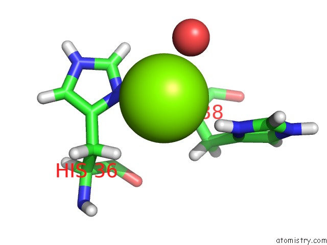
Mono view
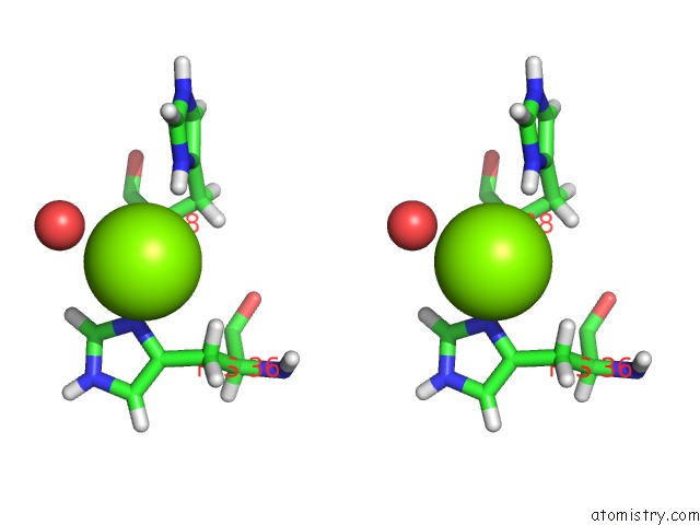
Stereo pair view
|
A full contact list of Magnesium with other atoms in the Mg binding
site number 4 of High Resolution Structure of the Catalytic Domain of Mannanase SACTE_2347 From Streptomyces Sp. Sirexaa-E within 5.0Å range:
probe
|
atom
|
residue
|
distance (Å)
|
B
|
Occ
|
A:Mg405
b:26.1
occ:1.00
|
HD1
|
A:HIS36
|
1.9
|
22.8
|
0.5
|
HD1
|
A:HIS38
|
2.4
|
20.1
|
1.0
|
ND1
|
A:HIS36
|
2.5
|
19.0
|
0.5
|
HA
|
A:HIS38
|
2.7
|
21.6
|
1.0
|
HB3
|
A:HIS36
|
3.0
|
24.1
|
0.5
|
O
|
A:HOH597
|
3.0
|
15.6
|
1.0
|
ND1
|
A:HIS38
|
3.2
|
16.8
|
1.0
|
CE1
|
A:HIS36
|
3.3
|
18.8
|
0.5
|
CG
|
A:HIS36
|
3.3
|
19.5
|
0.5
|
HE1
|
A:HIS36
|
3.5
|
22.5
|
0.5
|
HB2
|
A:HIS38
|
3.6
|
20.5
|
1.0
|
H
|
A:HIS38
|
3.6
|
22.6
|
0.5
|
CA
|
A:HIS38
|
3.6
|
18.0
|
1.0
|
CB
|
A:HIS36
|
3.6
|
20.1
|
0.5
|
CB
|
A:HIS38
|
3.9
|
17.1
|
1.0
|
CG
|
A:HIS38
|
4.0
|
16.4
|
1.0
|
N
|
A:HIS38
|
4.0
|
18.8
|
1.0
|
HB2
|
A:HIS36
|
4.1
|
24.1
|
0.5
|
CE1
|
A:HIS38
|
4.3
|
14.9
|
1.0
|
NE2
|
A:HIS36
|
4.3
|
18.9
|
0.5
|
CD2
|
A:HIS36
|
4.3
|
19.6
|
0.5
|
HE1
|
A:HIS38
|
4.4
|
17.9
|
1.0
|
C
|
A:HIS38
|
4.7
|
17.9
|
1.0
|
HB3
|
A:HIS38
|
4.8
|
20.5
|
1.0
|
CA
|
A:HIS36
|
4.9
|
20.9
|
0.5
|
C
|
A:HIS36
|
4.9
|
20.0
|
0.5
|
O
|
A:HIS36
|
4.9
|
19.9
|
0.5
|
|
Magnesium binding site 5 out
of 5 in 4fk9
Go back to
Magnesium Binding Sites List in 4fk9
Magnesium binding site 5 out
of 5 in the High Resolution Structure of the Catalytic Domain of Mannanase SACTE_2347 From Streptomyces Sp. Sirexaa-E
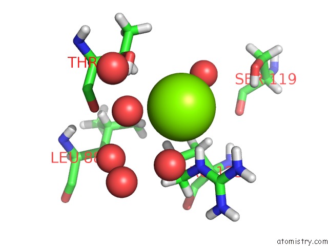
Mono view
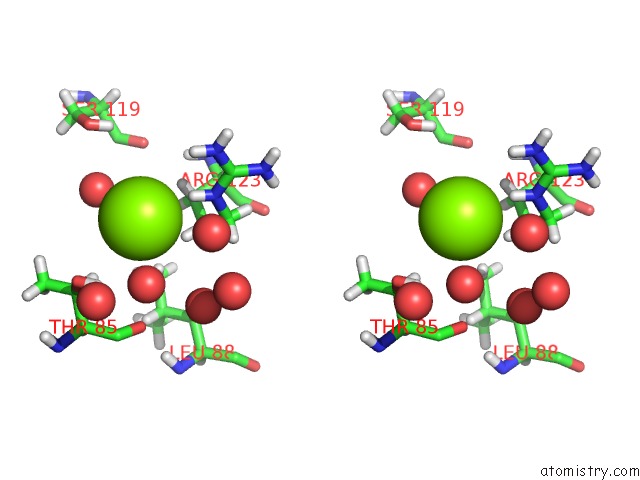
Stereo pair view
|
A full contact list of Magnesium with other atoms in the Mg binding
site number 5 of High Resolution Structure of the Catalytic Domain of Mannanase SACTE_2347 From Streptomyces Sp. Sirexaa-E within 5.0Å range:
probe
|
atom
|
residue
|
distance (Å)
|
B
|
Occ
|
A:Mg407
b:13.4
occ:1.00
|
HE
|
A:ARG123
|
2.4
|
8.9
|
1.0
|
O
|
A:HOH786
|
3.0
|
22.3
|
1.0
|
HG2
|
A:ARG123
|
3.1
|
8.2
|
1.0
|
NE
|
A:ARG123
|
3.3
|
7.4
|
1.0
|
HG1
|
A:THR85
|
3.3
|
16.0
|
1.0
|
O
|
A:HOH528
|
3.4
|
9.7
|
1.0
|
O
|
A:HOH997
|
3.5
|
31.1
|
1.0
|
HB
|
A:THR85
|
3.5
|
11.0
|
1.0
|
HH11
|
A:ARG123
|
3.6
|
9.9
|
1.0
|
OG1
|
A:THR85
|
3.6
|
13.3
|
1.0
|
HB2
|
A:ARG123
|
3.7
|
7.6
|
1.0
|
CG
|
A:ARG123
|
3.8
|
6.8
|
1.0
|
CD
|
A:ARG123
|
4.0
|
7.0
|
1.0
|
HD2
|
A:ARG123
|
4.1
|
8.4
|
1.0
|
CB
|
A:THR85
|
4.1
|
9.2
|
1.0
|
CZ
|
A:ARG123
|
4.2
|
7.3
|
1.0
|
NH1
|
A:ARG123
|
4.2
|
8.2
|
1.0
|
CB
|
A:ARG123
|
4.3
|
6.3
|
1.0
|
O
|
A:HOH568
|
4.6
|
10.2
|
1.0
|
O
|
A:HOH907
|
4.6
|
39.7
|
0.5
|
HB3
|
A:ARG123
|
4.6
|
7.6
|
1.0
|
HD12
|
A:LEU88
|
4.6
|
10.7
|
1.0
|
HG3
|
A:ARG123
|
4.7
|
8.2
|
1.0
|
O
|
A:THR85
|
4.8
|
8.0
|
1.0
|
HG22
|
A:THR85
|
4.8
|
12.6
|
1.0
|
HD3
|
A:ARG123
|
4.9
|
8.4
|
1.0
|
HG
|
A:SER119
|
4.9
|
7.6
|
1.0
|
O
|
A:HOH656
|
4.9
|
18.4
|
1.0
|
C
|
A:THR85
|
5.0
|
7.4
|
1.0
|
HH12
|
A:ARG123
|
5.0
|
9.9
|
1.0
|
|
Reference:
T.E.Takasuka,
J.F.Acheson,
C.M.Bianchetti,
B.M.Prom,
L.F.Bergeman,
A.J.Book,
C.R.Currie,
B.G.Fox.
Biochemical Properties and Atomic Resolution Structure of A Proteolytically Processed Beta-Mannanase From Cellulolytic Streptomyces Sp. Sirexaa-E. Plos One V. 9 94166 2014.
ISSN: ESSN 1932-6203
PubMed: 24710170
DOI: 10.1371/JOURNAL.PONE.0094166
Page generated: Fri Aug 16 15:09:11 2024
|