Magnesium »
PDB 4fk1-4fs5 »
4fmo »
Magnesium in PDB 4fmo: Structure of the C-Terminal Domain of the Saccharomyces Cerevisiae Mutl Alpha (MLH1/PMS1) Heterodimer Bound to A Fragment of EXO1
Protein crystallography data
The structure of Structure of the C-Terminal Domain of the Saccharomyces Cerevisiae Mutl Alpha (MLH1/PMS1) Heterodimer Bound to A Fragment of EXO1, PDB code: 4fmo
was solved by
E.Gueneau,
P.Legrand,
J.B.Charbonnier,
with X-Ray Crystallography technique. A brief refinement statistics is given in the table below:
| Resolution Low / High (Å) | 48.41 / 3.04 |
| Space group | C 1 2 1 |
| Cell size a, b, c (Å), α, β, γ (°) | 193.700, 66.140, 74.470, 90.00, 91.28, 90.00 |
| R / Rfree (%) | 18 / 19.6 |
Other elements in 4fmo:
The structure of Structure of the C-Terminal Domain of the Saccharomyces Cerevisiae Mutl Alpha (MLH1/PMS1) Heterodimer Bound to A Fragment of EXO1 also contains other interesting chemical elements:
| Zinc | (Zn) | 2 atoms |
Magnesium Binding Sites:
The binding sites of Magnesium atom in the Structure of the C-Terminal Domain of the Saccharomyces Cerevisiae Mutl Alpha (MLH1/PMS1) Heterodimer Bound to A Fragment of EXO1
(pdb code 4fmo). This binding sites where shown within
5.0 Angstroms radius around Magnesium atom.
In total only one binding site of Magnesium was determined in the Structure of the C-Terminal Domain of the Saccharomyces Cerevisiae Mutl Alpha (MLH1/PMS1) Heterodimer Bound to A Fragment of EXO1, PDB code: 4fmo:
In total only one binding site of Magnesium was determined in the Structure of the C-Terminal Domain of the Saccharomyces Cerevisiae Mutl Alpha (MLH1/PMS1) Heterodimer Bound to A Fragment of EXO1, PDB code: 4fmo:
Magnesium binding site 1 out of 1 in 4fmo
Go back to
Magnesium binding site 1 out
of 1 in the Structure of the C-Terminal Domain of the Saccharomyces Cerevisiae Mutl Alpha (MLH1/PMS1) Heterodimer Bound to A Fragment of EXO1
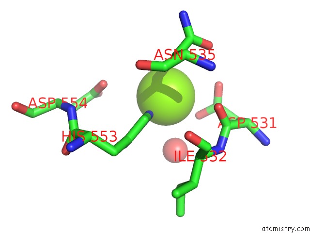
Mono view
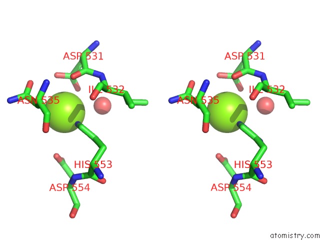
Stereo pair view
Mono view
Stereo pair view
A full contact list of Magnesium with other atoms in the Mg binding
site number 1 of Structure of the C-Terminal Domain of the Saccharomyces Cerevisiae Mutl Alpha (MLH1/PMS1) Heterodimer Bound to A Fragment of EXO1 within 5.0Å range:
|
Reference:
E.Gueneau,
C.Dherin,
P.Legrand,
C.Tellier-Lebegue,
B.Gilquin,
P.Bonnesoeur,
F.Londino,
C.Quemener,
M.H.Le Du,
J.A.Marquez,
M.Moutiez,
M.Gondry,
S.Boiteux,
J.B.Charbonnier.
Structure of the Mutl Alpha C-Terminal Domain Reveals How MLH1 Contributes to PMS1 Endonuclease Site. Nat.Struct.Mol.Biol. V. 20 461 2013.
ISSN: ISSN 1545-9993
PubMed: 23435383
DOI: 10.1038/NSMB.2511
Page generated: Fri Aug 16 15:12:48 2024
ISSN: ISSN 1545-9993
PubMed: 23435383
DOI: 10.1038/NSMB.2511
Last articles
Fe in 2YXOFe in 2YRS
Fe in 2YXC
Fe in 2YNM
Fe in 2YVJ
Fe in 2YP1
Fe in 2YU2
Fe in 2YU1
Fe in 2YQB
Fe in 2YOO