Magnesium »
PDB 4fut-4g7h »
4fvr »
Magnesium in PDB 4fvr: Crystal Structure of the JAK2 Pseudokinase Domain Mutant V617F (Mg- Atp-Bound Form)
Enzymatic activity of Crystal Structure of the JAK2 Pseudokinase Domain Mutant V617F (Mg- Atp-Bound Form)
All present enzymatic activity of Crystal Structure of the JAK2 Pseudokinase Domain Mutant V617F (Mg- Atp-Bound Form):
2.7.10.2;
2.7.10.2;
Protein crystallography data
The structure of Crystal Structure of the JAK2 Pseudokinase Domain Mutant V617F (Mg- Atp-Bound Form), PDB code: 4fvr
was solved by
R.M.Bandaranayake,
S.R.Hubbard,
with X-Ray Crystallography technique. A brief refinement statistics is given in the table below:
| Resolution Low / High (Å) | 43.50 / 2.00 |
| Space group | P 1 21 1 |
| Cell size a, b, c (Å), α, β, γ (°) | 46.900, 57.326, 60.555, 90.00, 111.86, 90.00 |
| R / Rfree (%) | 18.1 / 22.9 |
Magnesium Binding Sites:
The binding sites of Magnesium atom in the Crystal Structure of the JAK2 Pseudokinase Domain Mutant V617F (Mg- Atp-Bound Form)
(pdb code 4fvr). This binding sites where shown within
5.0 Angstroms radius around Magnesium atom.
In total only one binding site of Magnesium was determined in the Crystal Structure of the JAK2 Pseudokinase Domain Mutant V617F (Mg- Atp-Bound Form), PDB code: 4fvr:
In total only one binding site of Magnesium was determined in the Crystal Structure of the JAK2 Pseudokinase Domain Mutant V617F (Mg- Atp-Bound Form), PDB code: 4fvr:
Magnesium binding site 1 out of 1 in 4fvr
Go back to
Magnesium binding site 1 out
of 1 in the Crystal Structure of the JAK2 Pseudokinase Domain Mutant V617F (Mg- Atp-Bound Form)
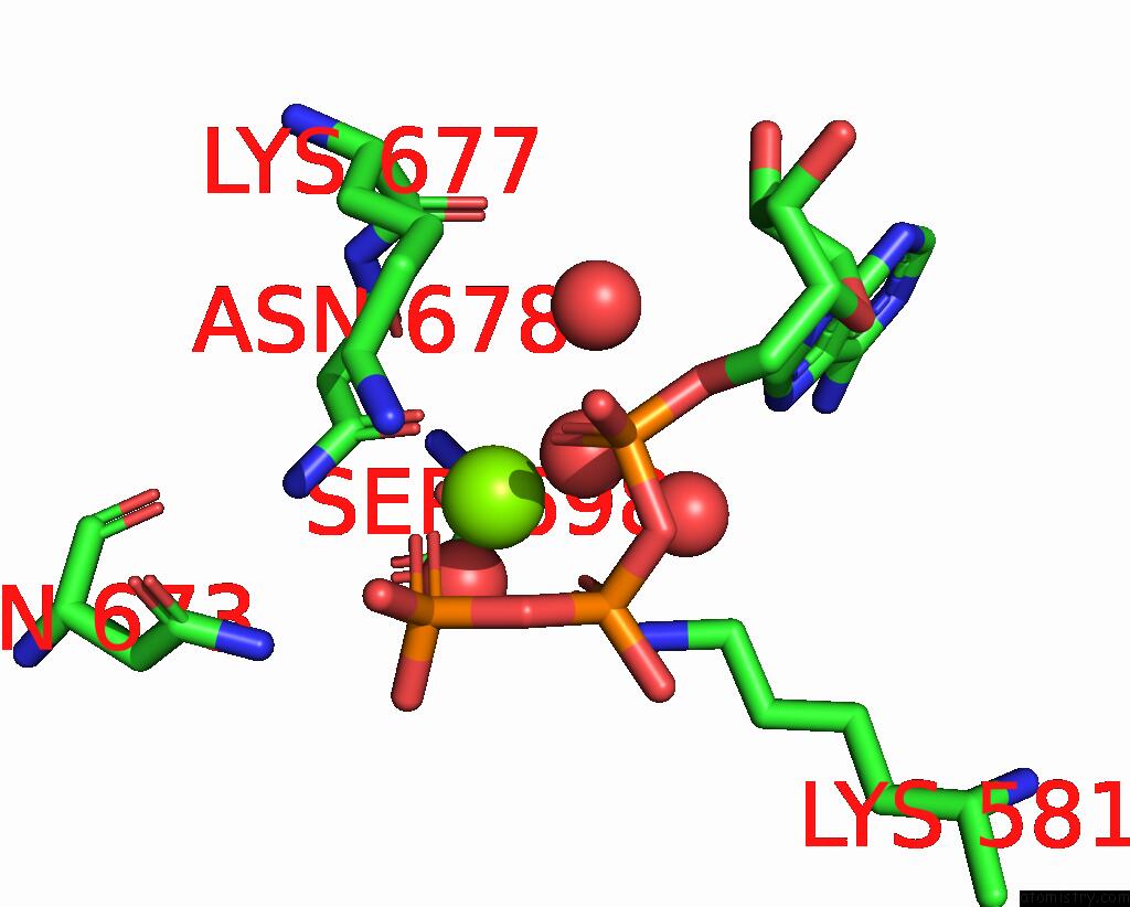
Mono view
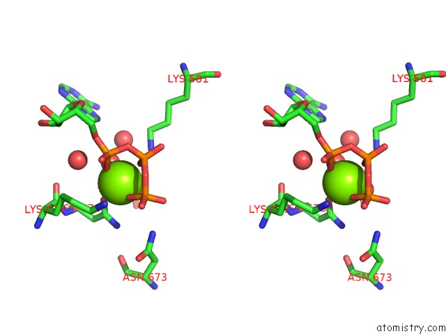
Stereo pair view
Mono view
Stereo pair view
A full contact list of Magnesium with other atoms in the Mg binding
site number 1 of Crystal Structure of the JAK2 Pseudokinase Domain Mutant V617F (Mg- Atp-Bound Form) within 5.0Å range:
|
Reference:
R.M.Bandaranayake,
D.Ungureanu,
Y.Shan,
D.E.Shaw,
O.Silvennoinen,
S.R.Hubbard.
Crystal Structures of the JAK2 Pseudokinase Domain and the Pathogenic Mutant V617F. Nat.Struct.Mol.Biol. V. 19 754 2012.
ISSN: ISSN 1545-9993
PubMed: 22820988
DOI: 10.1038/NSMB.2348
Page generated: Mon Aug 11 13:04:21 2025
ISSN: ISSN 1545-9993
PubMed: 22820988
DOI: 10.1038/NSMB.2348
Last articles
Mg in 5DH9Mg in 5DH4
Mg in 5DGH
Mg in 5DGT
Mg in 5DGK
Mg in 5DEI
Mg in 5DGI
Mg in 5DGD
Mg in 5DGB
Mg in 5DGA