Magnesium »
PDB 4fut-4g7h »
4g7f »
Magnesium in PDB 4g7f: Crystal Structure of Enolase From Trypanosoma Cruzi
Enzymatic activity of Crystal Structure of Enolase From Trypanosoma Cruzi
All present enzymatic activity of Crystal Structure of Enolase From Trypanosoma Cruzi:
4.2.1.11;
4.2.1.11;
Protein crystallography data
The structure of Crystal Structure of Enolase From Trypanosoma Cruzi, PDB code: 4g7f
was solved by
T.K.Craig,
T.E.Edwards,
B.Staker,
Seattle Structural Genomics Center Forinfectious Disease (Ssgcid),
with X-Ray Crystallography technique. A brief refinement statistics is given in the table below:
| Resolution Low / High (Å) | 19.81 / 2.40 |
| Space group | C 2 2 21 |
| Cell size a, b, c (Å), α, β, γ (°) | 75.290, 119.310, 110.360, 90.00, 90.00, 90.00 |
| R / Rfree (%) | 18.9 / 25 |
Magnesium Binding Sites:
The binding sites of Magnesium atom in the Crystal Structure of Enolase From Trypanosoma Cruzi
(pdb code 4g7f). This binding sites where shown within
5.0 Angstroms radius around Magnesium atom.
In total only one binding site of Magnesium was determined in the Crystal Structure of Enolase From Trypanosoma Cruzi, PDB code: 4g7f:
In total only one binding site of Magnesium was determined in the Crystal Structure of Enolase From Trypanosoma Cruzi, PDB code: 4g7f:
Magnesium binding site 1 out of 1 in 4g7f
Go back to
Magnesium binding site 1 out
of 1 in the Crystal Structure of Enolase From Trypanosoma Cruzi
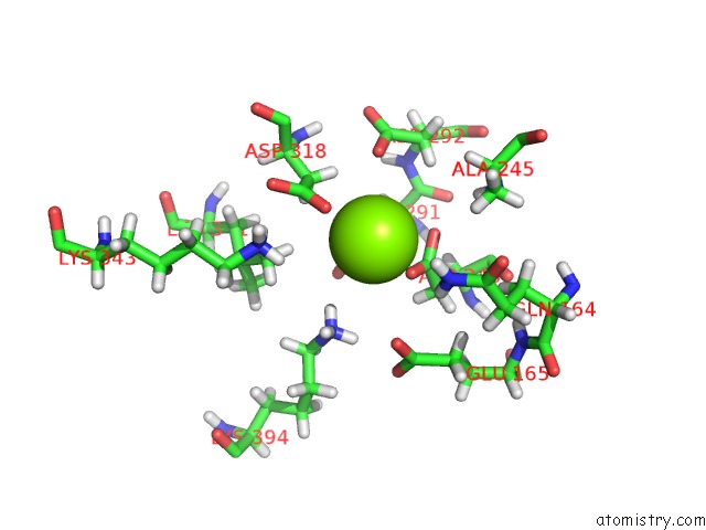
Mono view
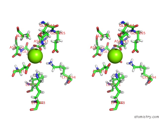
Stereo pair view
Mono view
Stereo pair view
A full contact list of Magnesium with other atoms in the Mg binding
site number 1 of Crystal Structure of Enolase From Trypanosoma Cruzi within 5.0Å range:
|
Reference:
T.K.Craig,
T.E.Edwards,
B.Staker,
Seattle Structural Genomics Center For Infectious Disease(Ssgcid).
Crystal Structure of Enolase From Trypanosoma Cruzi To Be Published.
Page generated: Mon Aug 11 13:10:23 2025
Last articles
Mg in 5SC8Mg in 5SCA
Mg in 5SBE
Mg in 5SBD
Mg in 5SBC
Mg in 5SBB
Mg in 5SBA
Mg in 5SB8
Mg in 5SB9
Mg in 5SB7