Magnesium »
PDB 4g7h-4gni »
4g87 »
Magnesium in PDB 4g87: Crystal Structure of Glmu From Mycobacterium Tuberculosis Snapshot 1
Enzymatic activity of Crystal Structure of Glmu From Mycobacterium Tuberculosis Snapshot 1
All present enzymatic activity of Crystal Structure of Glmu From Mycobacterium Tuberculosis Snapshot 1:
2.3.1.157; 2.7.7.23;
2.3.1.157; 2.7.7.23;
Protein crystallography data
The structure of Crystal Structure of Glmu From Mycobacterium Tuberculosis Snapshot 1, PDB code: 4g87
was solved by
P.A.Jagtap,
S.K.Verma,
B.Prakash,
with X-Ray Crystallography technique. A brief refinement statistics is given in the table below:
| Resolution Low / High (Å) | 18.84 / 2.03 |
| Space group | H 3 |
| Cell size a, b, c (Å), α, β, γ (°) | 77.000, 77.000, 276.730, 90.00, 90.00, 120.00 |
| R / Rfree (%) | 17.8 / 23.5 |
Other elements in 4g87:
The structure of Crystal Structure of Glmu From Mycobacterium Tuberculosis Snapshot 1 also contains other interesting chemical elements:
| Cobalt | (Co) | 2 atoms |
Magnesium Binding Sites:
The binding sites of Magnesium atom in the Crystal Structure of Glmu From Mycobacterium Tuberculosis Snapshot 1
(pdb code 4g87). This binding sites where shown within
5.0 Angstroms radius around Magnesium atom.
In total 2 binding sites of Magnesium where determined in the Crystal Structure of Glmu From Mycobacterium Tuberculosis Snapshot 1, PDB code: 4g87:
Jump to Magnesium binding site number: 1; 2;
In total 2 binding sites of Magnesium where determined in the Crystal Structure of Glmu From Mycobacterium Tuberculosis Snapshot 1, PDB code: 4g87:
Jump to Magnesium binding site number: 1; 2;
Magnesium binding site 1 out of 2 in 4g87
Go back to
Magnesium binding site 1 out
of 2 in the Crystal Structure of Glmu From Mycobacterium Tuberculosis Snapshot 1
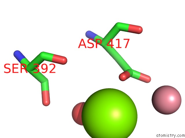
Mono view
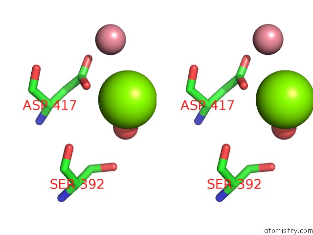
Stereo pair view
Mono view
Stereo pair view
A full contact list of Magnesium with other atoms in the Mg binding
site number 1 of Crystal Structure of Glmu From Mycobacterium Tuberculosis Snapshot 1 within 5.0Å range:
|
Magnesium binding site 2 out of 2 in 4g87
Go back to
Magnesium binding site 2 out
of 2 in the Crystal Structure of Glmu From Mycobacterium Tuberculosis Snapshot 1
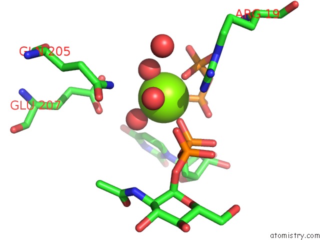
Mono view
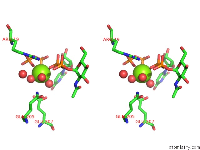
Stereo pair view
Mono view
Stereo pair view
A full contact list of Magnesium with other atoms in the Mg binding
site number 2 of Crystal Structure of Glmu From Mycobacterium Tuberculosis Snapshot 1 within 5.0Å range:
|
Reference:
P.K.A.Jagtap,
S.K.Verma,
N.Vithani,
V.S.Bais,
B.Prakash.
Crystal Structures Identify An Atypical Two-Metal-Ion Mechanism For Uridyltransfer in Glmu: Its Significance to Sugar Nucleotidyl Transferases J.Mol.Biol. V. 425 1745 2013.
ISSN: ISSN 0022-2836
PubMed: 23485416
DOI: 10.1016/J.JMB.2013.02.019
Page generated: Fri Aug 16 15:27:34 2024
ISSN: ISSN 0022-2836
PubMed: 23485416
DOI: 10.1016/J.JMB.2013.02.019
Last articles
Zn in 9J0NZn in 9J0O
Zn in 9J0P
Zn in 9FJX
Zn in 9EKB
Zn in 9C0F
Zn in 9CAH
Zn in 9CH0
Zn in 9CH3
Zn in 9CH1