Magnesium »
PDB 4g7h-4gni »
4gbf »
Magnesium in PDB 4gbf: Crystal Structure of the C-Terminal Domain of GP131 From Bacteriophage Phikz
Protein crystallography data
The structure of Crystal Structure of the C-Terminal Domain of GP131 From Bacteriophage Phikz, PDB code: 4gbf
was solved by
L.V.Sycheva,
M.M.Shneider,
P.G.Leiman,
with X-Ray Crystallography technique. A brief refinement statistics is given in the table below:
| Resolution Low / High (Å) | 43.18 / 1.95 |
| Space group | P 21 21 21 |
| Cell size a, b, c (Å), α, β, γ (°) | 79.449, 86.353, 97.362, 90.00, 90.00, 90.00 |
| R / Rfree (%) | 16.1 / 19.9 |
Other elements in 4gbf:
The structure of Crystal Structure of the C-Terminal Domain of GP131 From Bacteriophage Phikz also contains other interesting chemical elements:
| Chlorine | (Cl) | 4 atoms |
Magnesium Binding Sites:
The binding sites of Magnesium atom in the Crystal Structure of the C-Terminal Domain of GP131 From Bacteriophage Phikz
(pdb code 4gbf). This binding sites where shown within
5.0 Angstroms radius around Magnesium atom.
In total 5 binding sites of Magnesium where determined in the Crystal Structure of the C-Terminal Domain of GP131 From Bacteriophage Phikz, PDB code: 4gbf:
Jump to Magnesium binding site number: 1; 2; 3; 4; 5;
In total 5 binding sites of Magnesium where determined in the Crystal Structure of the C-Terminal Domain of GP131 From Bacteriophage Phikz, PDB code: 4gbf:
Jump to Magnesium binding site number: 1; 2; 3; 4; 5;
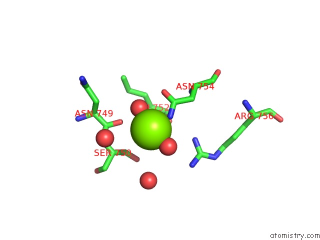
Magnesium binding site 1 out of 5 in 4gbf
Go back to
Magnesium binding site 1 out
of 5 in the Crystal Structure of the C-Terminal Domain of GP131 From Bacteriophage Phikz

Mono view

Stereo pair view
Mono view
Stereo pair view
A full contact list of Magnesium with other atoms in the Mg binding
site number 1 of Crystal Structure of the C-Terminal Domain of GP131 From Bacteriophage Phikz within 5.0Å range:
|
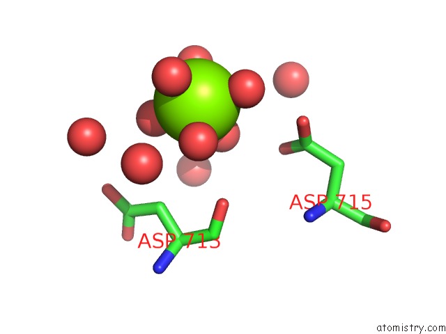
Magnesium binding site 2 out of 5 in 4gbf
Go back to
Magnesium binding site 2 out
of 5 in the Crystal Structure of the C-Terminal Domain of GP131 From Bacteriophage Phikz
Mono view

Stereo pair view
Mono view
Stereo pair view
A full contact list of Magnesium with other atoms in the Mg binding
site number 2 of Crystal Structure of the C-Terminal Domain of GP131 From Bacteriophage Phikz within 5.0Å range:
|
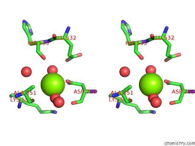
Magnesium binding site 3 out of 5 in 4gbf
Go back to
Magnesium binding site 3 out
of 5 in the Crystal Structure of the C-Terminal Domain of GP131 From Bacteriophage Phikz
Mono view

Stereo pair view
Mono view
Stereo pair view
A full contact list of Magnesium with other atoms in the Mg binding
site number 3 of Crystal Structure of the C-Terminal Domain of GP131 From Bacteriophage Phikz within 5.0Å range:
|
Magnesium binding site 4 out of 5 in 4gbf
Go back to
Magnesium binding site 4 out
of 5 in the Crystal Structure of the C-Terminal Domain of GP131 From Bacteriophage Phikz

Mono view

Stereo pair view
Mono view
Stereo pair view
A full contact list of Magnesium with other atoms in the Mg binding
site number 4 of Crystal Structure of the C-Terminal Domain of GP131 From Bacteriophage Phikz within 5.0Å range:
|
Magnesium binding site 5 out of 5 in 4gbf
Go back to
Magnesium binding site 5 out
of 5 in the Crystal Structure of the C-Terminal Domain of GP131 From Bacteriophage Phikz

Mono view
Stereo pair view
Mono view
Stereo pair view
A full contact list of Magnesium with other atoms in the Mg binding
site number 5 of Crystal Structure of the C-Terminal Domain of GP131 From Bacteriophage Phikz within 5.0Å range:
|
Reference:
L.V.Sycheva,
M.M.Shneider,
N.N.Sykilinda,
M.A.Ivanova,
K.A.Miroshnikov,
P.G.Leiman.
Crystal Structure and Location of GP131 in the Bacteriophage Phikz Virion. Virology V. 434 257 2012.
ISSN: ISSN 0042-6822
PubMed: 23031178
DOI: 10.1016/J.VIROL.2012.09.001
Page generated: Fri Aug 16 15:28:56 2024
ISSN: ISSN 0042-6822
PubMed: 23031178
DOI: 10.1016/J.VIROL.2012.09.001
Last articles
Zn in 9J0NZn in 9J0O
Zn in 9J0P
Zn in 9FJX
Zn in 9EKB
Zn in 9C0F
Zn in 9CAH
Zn in 9CH0
Zn in 9CH3
Zn in 9CH1