Magnesium »
PDB 4gnr-4gyq »
4gt8 »
Magnesium in PDB 4gt8: Crystal Structure of the Catalytic and Atp-Binding Domain From Vras in Complex with Adp
Enzymatic activity of Crystal Structure of the Catalytic and Atp-Binding Domain From Vras in Complex with Adp
All present enzymatic activity of Crystal Structure of the Catalytic and Atp-Binding Domain From Vras in Complex with Adp:
2.7.13.3;
2.7.13.3;
Protein crystallography data
The structure of Crystal Structure of the Catalytic and Atp-Binding Domain From Vras in Complex with Adp, PDB code: 4gt8
was solved by
P.G.Leonard,
J.Valverde,
A.M.Stock,
with X-Ray Crystallography technique. A brief refinement statistics is given in the table below:
| Resolution Low / High (Å) | 29.45 / 1.51 |
| Space group | P 21 21 21 |
| Cell size a, b, c (Å), α, β, γ (°) | 31.192, 47.233, 89.398, 90.00, 90.00, 90.00 |
| R / Rfree (%) | 18.3 / 19.9 |
Magnesium Binding Sites:
The binding sites of Magnesium atom in the Crystal Structure of the Catalytic and Atp-Binding Domain From Vras in Complex with Adp
(pdb code 4gt8). This binding sites where shown within
5.0 Angstroms radius around Magnesium atom.
In total only one binding site of Magnesium was determined in the Crystal Structure of the Catalytic and Atp-Binding Domain From Vras in Complex with Adp, PDB code: 4gt8:
In total only one binding site of Magnesium was determined in the Crystal Structure of the Catalytic and Atp-Binding Domain From Vras in Complex with Adp, PDB code: 4gt8:
Magnesium binding site 1 out of 1 in 4gt8
Go back to
Magnesium binding site 1 out
of 1 in the Crystal Structure of the Catalytic and Atp-Binding Domain From Vras in Complex with Adp
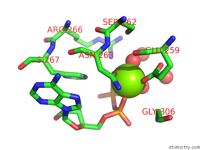
Mono view
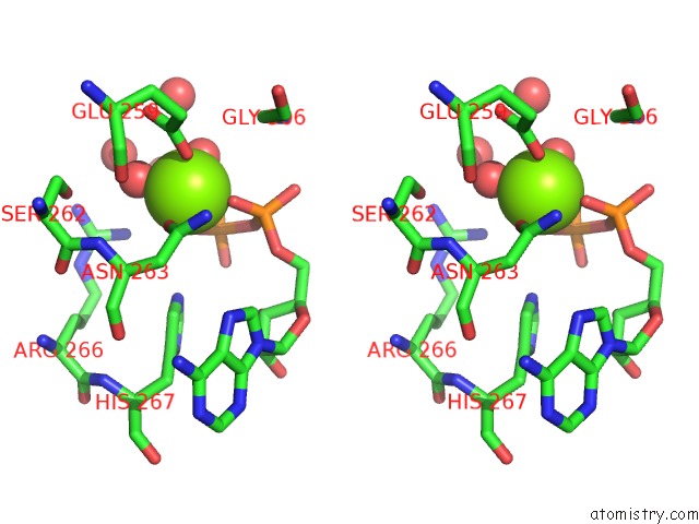
Stereo pair view
Mono view
Stereo pair view
A full contact list of Magnesium with other atoms in the Mg binding
site number 1 of Crystal Structure of the Catalytic and Atp-Binding Domain From Vras in Complex with Adp within 5.0Å range:
|
Reference:
P.G.Leonard,
J.Valverde,
A.M.Stock.
Structure of the Staphylococcus Aureus Vras Catalytic and Atp-Binding Domain To Be Published.
Page generated: Mon Aug 11 13:37:36 2025
Last articles
Mg in 5JJLMg in 5JJK
Mg in 5JJI
Mg in 5JJS
Mg in 5JJR
Mg in 5JHS
Mg in 5JJG
Mg in 5JHR
Mg in 5JJB
Mg in 5JIP