Magnesium »
PDB 4gnr-4gyq »
4gue »
Magnesium in PDB 4gue: Structure of N-Terminal Kinase Domain of RSK2 with Flavonoid Glycoside Quercitrin
Enzymatic activity of Structure of N-Terminal Kinase Domain of RSK2 with Flavonoid Glycoside Quercitrin
All present enzymatic activity of Structure of N-Terminal Kinase Domain of RSK2 with Flavonoid Glycoside Quercitrin:
2.7.11.1;
2.7.11.1;
Protein crystallography data
The structure of Structure of N-Terminal Kinase Domain of RSK2 with Flavonoid Glycoside Quercitrin, PDB code: 4gue
was solved by
U.Derewenda,
D.Utepbergenov,
G.Szukalska,
Z.S.Derewenda,
with X-Ray Crystallography technique. A brief refinement statistics is given in the table below:
| Resolution Low / High (Å) | 25.29 / 1.80 |
| Space group | C 1 2 1 |
| Cell size a, b, c (Å), α, β, γ (°) | 98.084, 40.686, 83.264, 90.00, 114.31, 90.00 |
| R / Rfree (%) | 17.1 / 20.5 |
Magnesium Binding Sites:
The binding sites of Magnesium atom in the Structure of N-Terminal Kinase Domain of RSK2 with Flavonoid Glycoside Quercitrin
(pdb code 4gue). This binding sites where shown within
5.0 Angstroms radius around Magnesium atom.
In total only one binding site of Magnesium was determined in the Structure of N-Terminal Kinase Domain of RSK2 with Flavonoid Glycoside Quercitrin, PDB code: 4gue:
In total only one binding site of Magnesium was determined in the Structure of N-Terminal Kinase Domain of RSK2 with Flavonoid Glycoside Quercitrin, PDB code: 4gue:
Magnesium binding site 1 out of 1 in 4gue
Go back to
Magnesium binding site 1 out
of 1 in the Structure of N-Terminal Kinase Domain of RSK2 with Flavonoid Glycoside Quercitrin
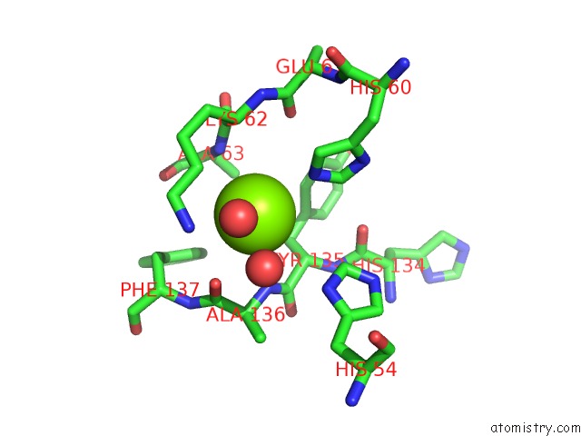
Mono view
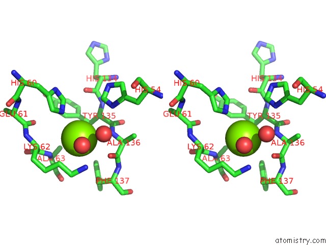
Stereo pair view
Mono view
Stereo pair view
A full contact list of Magnesium with other atoms in the Mg binding
site number 1 of Structure of N-Terminal Kinase Domain of RSK2 with Flavonoid Glycoside Quercitrin within 5.0Å range:
|
Reference:
U.Derewenda,
M.Artamonov,
G.Szukalska,
D.Utepbergenov,
N.Olekhnovich,
H.I.Parikh,
G.E.Kellogg,
A.V.Somlyo,
Z.S.Derewenda.
Identification of Quercitrin As An Inhibitor of the P90 S6 Ribosomal Kinase (Rsk): Structure of Its Complex with the N-Terminal Domain of RSK2 at 1.8 A Resolution. Acta Crystallogr.,Sect.D V. 69 266 2013.
ISSN: ISSN 0907-4449
PubMed: 23385462
DOI: 10.1107/S0907444912045520
Page generated: Mon Aug 11 13:37:54 2025
ISSN: ISSN 0907-4449
PubMed: 23385462
DOI: 10.1107/S0907444912045520
Last articles
Mg in 5FPHMg in 5FR2
Mg in 5FQ8
Mg in 5FR1
Mg in 5FQO
Mg in 5FQ7
Mg in 5FQ5
Mg in 5FP3
Mg in 5FNV
Mg in 5FO5