Magnesium »
PDB 4gyz-4h9l »
4gzz »
Magnesium in PDB 4gzz: Crystal Structures of Bacterial Rna Polymerase Paused Elongation Complexes
Enzymatic activity of Crystal Structures of Bacterial Rna Polymerase Paused Elongation Complexes
All present enzymatic activity of Crystal Structures of Bacterial Rna Polymerase Paused Elongation Complexes:
2.7.7.6;
2.7.7.6;
Protein crystallography data
The structure of Crystal Structures of Bacterial Rna Polymerase Paused Elongation Complexes, PDB code: 4gzz
was solved by
A.Weixlbaumer,
K.Leon,
R.Landick,
S.A.Darst,
with X-Ray Crystallography technique. A brief refinement statistics is given in the table below:
| Resolution Low / High (Å) | 38.79 / 4.29 |
| Space group | H 3 |
| Cell size a, b, c (Å), α, β, γ (°) | 286.549, 286.549, 199.411, 90.00, 90.00, 120.00 |
| R / Rfree (%) | 23.5 / 28.5 |
Other elements in 4gzz:
The structure of Crystal Structures of Bacterial Rna Polymerase Paused Elongation Complexes also contains other interesting chemical elements:
| Zinc | (Zn) | 2 atoms |
Magnesium Binding Sites:
The binding sites of Magnesium atom in the Crystal Structures of Bacterial Rna Polymerase Paused Elongation Complexes
(pdb code 4gzz). This binding sites where shown within
5.0 Angstroms radius around Magnesium atom.
In total only one binding site of Magnesium was determined in the Crystal Structures of Bacterial Rna Polymerase Paused Elongation Complexes, PDB code: 4gzz:
In total only one binding site of Magnesium was determined in the Crystal Structures of Bacterial Rna Polymerase Paused Elongation Complexes, PDB code: 4gzz:
Magnesium binding site 1 out of 1 in 4gzz
Go back to
Magnesium binding site 1 out
of 1 in the Crystal Structures of Bacterial Rna Polymerase Paused Elongation Complexes
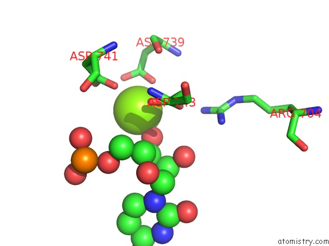
Mono view
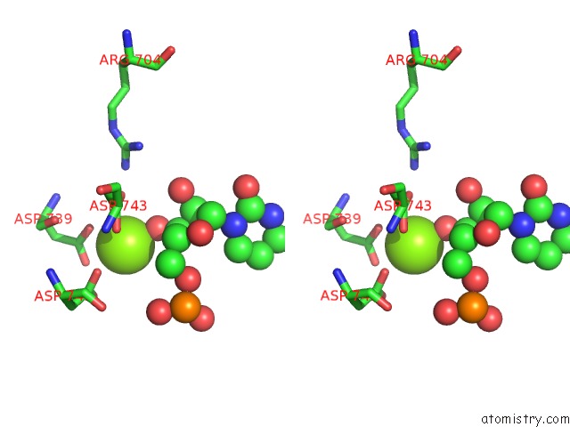
Stereo pair view
Mono view
Stereo pair view
A full contact list of Magnesium with other atoms in the Mg binding
site number 1 of Crystal Structures of Bacterial Rna Polymerase Paused Elongation Complexes within 5.0Å range:
|
Reference:
A.Weixlbaumer,
K.Leon,
R.Landick,
S.A.Darst.
Structural Basis of Transcriptional Pausing in Bacteria. Cell(Cambridge,Mass.) V. 152 431 2013.
ISSN: ISSN 0092-8674
PubMed: 23374340
DOI: 10.1016/J.CELL.2012.12.020
Page generated: Fri Aug 16 16:07:44 2024
ISSN: ISSN 0092-8674
PubMed: 23374340
DOI: 10.1016/J.CELL.2012.12.020
Last articles
Cl in 8AK8Cl in 8AK7
Cl in 8AJP
Cl in 8AJ6
Cl in 8AIO
Cl in 8AI7
Cl in 8AHZ
Cl in 8AHQ
Cl in 8AHY
Cl in 8AHO