Magnesium »
PDB 4hmy-4hyv »
4hnl »
Magnesium in PDB 4hnl: Crystal Structure of Enolase EGBG_01401 (Target Efi-502226) From Enterococcus Gallinarum EG2
Protein crystallography data
The structure of Crystal Structure of Enolase EGBG_01401 (Target Efi-502226) From Enterococcus Gallinarum EG2, PDB code: 4hnl
was solved by
Y.Patskovsky,
R.Toro,
R.Bhosle,
B.Hillerich,
R.D.Seidel,
E.Washington,
A.Scott Glenn,
S.Chowdhury,
B.Evans,
J.Hammonds,
W.D.Zencheck,
H.J.Imker,
N.F.Al Obaidi,
M.Stead,
J.Love,
J.A.Gerlt,
S.C.Almo,
Enzyme Functioninitiative (Efi),
with X-Ray Crystallography technique. A brief refinement statistics is given in the table below:
| Resolution Low / High (Å) | 47.64 / 1.48 |
| Space group | I 4 2 2 |
| Cell size a, b, c (Å), α, β, γ (°) | 115.943, 115.943, 119.970, 90.00, 90.00, 90.00 |
| R / Rfree (%) | 12.3 / 16.2 |
Other elements in 4hnl:
The structure of Crystal Structure of Enolase EGBG_01401 (Target Efi-502226) From Enterococcus Gallinarum EG2 also contains other interesting chemical elements:
| Chlorine | (Cl) | 2 atoms |
Magnesium Binding Sites:
The binding sites of Magnesium atom in the Crystal Structure of Enolase EGBG_01401 (Target Efi-502226) From Enterococcus Gallinarum EG2
(pdb code 4hnl). This binding sites where shown within
5.0 Angstroms radius around Magnesium atom.
In total only one binding site of Magnesium was determined in the Crystal Structure of Enolase EGBG_01401 (Target Efi-502226) From Enterococcus Gallinarum EG2, PDB code: 4hnl:
In total only one binding site of Magnesium was determined in the Crystal Structure of Enolase EGBG_01401 (Target Efi-502226) From Enterococcus Gallinarum EG2, PDB code: 4hnl:
Magnesium binding site 1 out of 1 in 4hnl
Go back to
Magnesium binding site 1 out
of 1 in the Crystal Structure of Enolase EGBG_01401 (Target Efi-502226) From Enterococcus Gallinarum EG2
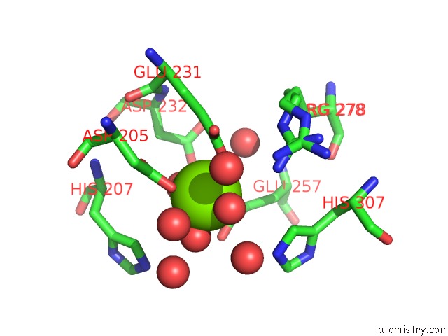
Mono view
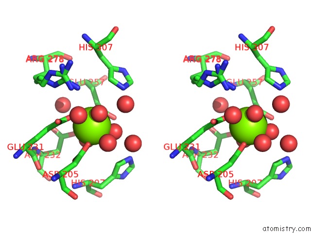
Stereo pair view
Mono view
Stereo pair view
A full contact list of Magnesium with other atoms in the Mg binding
site number 1 of Crystal Structure of Enolase EGBG_01401 (Target Efi-502226) From Enterococcus Gallinarum EG2 within 5.0Å range:
|
Reference:
Y.Patskovsky,
R.Toro,
R.Bhosle,
B.Hillerich,
R.D.Seidel,
E.Washington,
A.Scott Glenn,
S.Chowdhury,
B.Evans,
J.Hammonds,
W.D.Zencheck,
H.J.Imker,
N.F.Al Obaidi,
M.Stead,
J.Love,
J.A.Gerlt,
S.C.Almo.
Crystal Structure of Enolase EGBG_01401 From Enterococcus Gallinarum EG2 To Be Published.
Page generated: Mon Aug 11 13:53:34 2025
Last articles
Mg in 4LF4Mg in 4LF5
Mg in 4LCZ
Mg in 4LF2
Mg in 4LF1
Mg in 4LEM
Mg in 4LCK
Mg in 4LE0
Mg in 4LDZ
Mg in 4LDT