Magnesium »
PDB 4hmy-4hyv »
4ho7 »
Magnesium in PDB 4ho7: Crystal Structure of Eukaryotic Hslv From Trypanosoma Brucei
Protein crystallography data
The structure of Crystal Structure of Eukaryotic Hslv From Trypanosoma Brucei, PDB code: 4ho7
was solved by
K.H.Sung,
S.Y.Lee,
H.K.Song,
with X-Ray Crystallography technique. A brief refinement statistics is given in the table below:
| Resolution Low / High (Å) | 33.73 / 2.60 |
| Space group | I 2 2 2 |
| Cell size a, b, c (Å), α, β, γ (°) | 105.673, 111.062, 116.953, 90.00, 90.00, 90.00 |
| R / Rfree (%) | 21 / 24.8 |
Magnesium Binding Sites:
The binding sites of Magnesium atom in the Crystal Structure of Eukaryotic Hslv From Trypanosoma Brucei
(pdb code 4ho7). This binding sites where shown within
5.0 Angstroms radius around Magnesium atom.
In total 3 binding sites of Magnesium where determined in the Crystal Structure of Eukaryotic Hslv From Trypanosoma Brucei, PDB code: 4ho7:
Jump to Magnesium binding site number: 1; 2; 3;
In total 3 binding sites of Magnesium where determined in the Crystal Structure of Eukaryotic Hslv From Trypanosoma Brucei, PDB code: 4ho7:
Jump to Magnesium binding site number: 1; 2; 3;
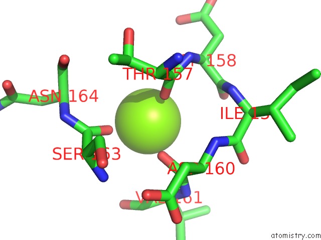
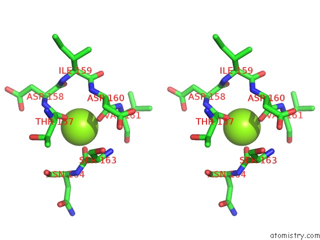
Magnesium binding site 1 out of 3 in 4ho7
Go back to
Magnesium binding site 1 out
of 3 in the Crystal Structure of Eukaryotic Hslv From Trypanosoma Brucei
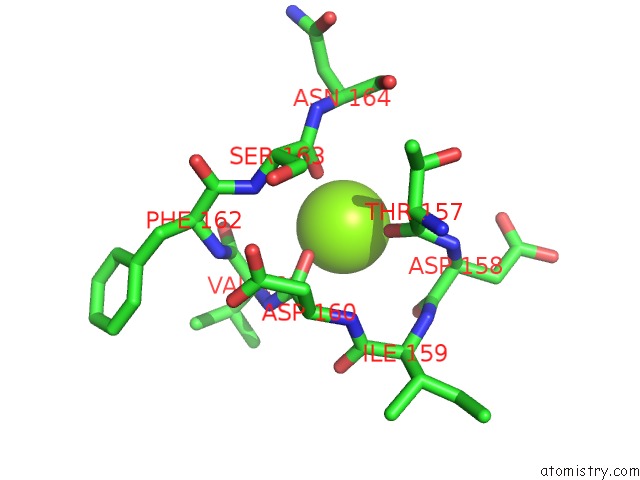
Mono view
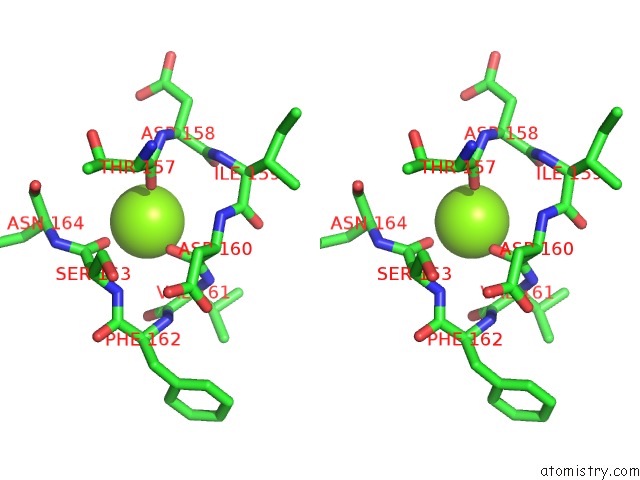
Stereo pair view
Mono view
Stereo pair view
A full contact list of Magnesium with other atoms in the Mg binding
site number 1 of Crystal Structure of Eukaryotic Hslv From Trypanosoma Brucei within 5.0Å range:
|
Magnesium binding site 2 out of 3 in 4ho7
Go back to
Magnesium binding site 2 out
of 3 in the Crystal Structure of Eukaryotic Hslv From Trypanosoma Brucei
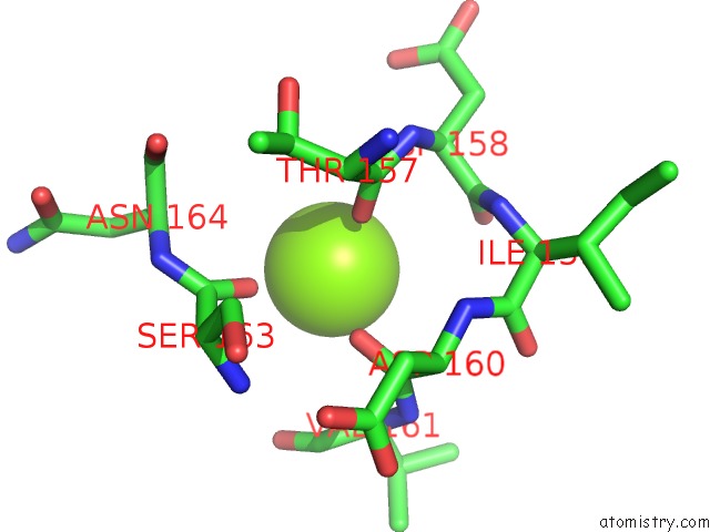
Mono view
Stereo pair view
Mono view
Stereo pair view
A full contact list of Magnesium with other atoms in the Mg binding
site number 2 of Crystal Structure of Eukaryotic Hslv From Trypanosoma Brucei within 5.0Å range:
|
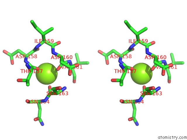
Magnesium binding site 3 out of 3 in 4ho7
Go back to
Magnesium binding site 3 out
of 3 in the Crystal Structure of Eukaryotic Hslv From Trypanosoma Brucei
Mono view
Stereo pair view
Mono view
Stereo pair view
A full contact list of Magnesium with other atoms in the Mg binding
site number 3 of Crystal Structure of Eukaryotic Hslv From Trypanosoma Brucei within 5.0Å range:
|
Reference:
K.H.Sung,
S.Y.Lee,
H.K.Song.
Structural and Biochemical Analyses of the Eukaryotic Heat Shock Locus V (Hslv) From Trypanosoma Brucei. J.Biol.Chem. V. 288 23234 2013.
ISSN: ISSN 0021-9258
PubMed: 23818520
DOI: 10.1074/JBC.M113.484832
Page generated: Mon Aug 11 13:54:06 2025
ISSN: ISSN 0021-9258
PubMed: 23818520
DOI: 10.1074/JBC.M113.484832
Last articles
Mg in 4LCZMg in 4LF2
Mg in 4LF1
Mg in 4LEM
Mg in 4LCK
Mg in 4LE0
Mg in 4LDZ
Mg in 4LDT
Mg in 4LA7
Mg in 4LDJ