Magnesium in PDB 4hqs: Crystal Structure of the Pneumoccocal Exposed Lipoprotein Thioredoxin SP_0659 (ETRX1) From Streptococcus Pneumoniae Strain TIGR4
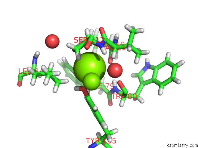
Protein crystallography data
The structure of Crystal Structure of the Pneumoccocal Exposed Lipoprotein Thioredoxin SP_0659 (ETRX1) From Streptococcus Pneumoniae Strain TIGR4, PDB code: 4hqs
was solved by
S.G.Bartual,
M.Saleh,
M.R.Abdullah,
I.Jensch,
T.M.Asmat,
L.Petruschka,
T.Pribyl,
J.A.Hermoso,
S.Hammerschmidt,
with X-Ray Crystallography technique. A brief refinement statistics is given in the table below:
Resolution Low / High (Å)
|
28.11 /
1.48
|
Space group
|
P 43 21 2
|
Cell size a, b, c (Å), α, β, γ (°)
|
62.850,
62.850,
89.600,
90.00,
90.00,
90.00
|
R / Rfree (%)
|
18.2 /
19.7
|
Magnesium Binding Sites:
The binding sites of Magnesium atom in the Crystal Structure of the Pneumoccocal Exposed Lipoprotein Thioredoxin SP_0659 (ETRX1) From Streptococcus Pneumoniae Strain TIGR4
(pdb code 4hqs). This binding sites where shown within
5.0 Angstroms radius around Magnesium atom.
In total 9 binding sites of Magnesium where determined in the
Crystal Structure of the Pneumoccocal Exposed Lipoprotein Thioredoxin SP_0659 (ETRX1) From Streptococcus Pneumoniae Strain TIGR4, PDB code: 4hqs:
Jump to Magnesium binding site number:
1;
2;
3;
4;
5;
6;
7;
8;
9;
Magnesium binding site 1 out
of 9 in 4hqs
Go back to
Magnesium Binding Sites List in 4hqs
Magnesium binding site 1 out
of 9 in the Crystal Structure of the Pneumoccocal Exposed Lipoprotein Thioredoxin SP_0659 (ETRX1) From Streptococcus Pneumoniae Strain TIGR4
Mono view
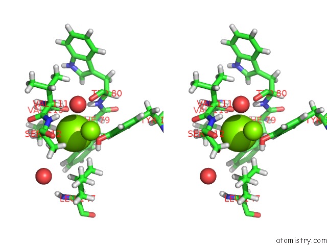
Stereo pair view
|
A full contact list of Magnesium with other atoms in the Mg binding
site number 1 of Crystal Structure of the Pneumoccocal Exposed Lipoprotein Thioredoxin SP_0659 (ETRX1) From Streptococcus Pneumoniae Strain TIGR4 within 5.0Å range:
probe
|
atom
|
residue
|
distance (Å)
|
B
|
Occ
|
A:Mg205
b:17.1
occ:1.00
|
HG
|
A:SER112
|
2.7
|
12.7
|
1.0
|
OG
|
A:SER112
|
2.8
|
10.6
|
1.0
|
HA
|
A:VAL111
|
2.8
|
9.3
|
1.0
|
HD22
|
A:LEU147
|
2.8
|
18.3
|
1.0
|
OH
|
A:TYR155
|
2.8
|
10.7
|
1.0
|
HD2
|
A:PHE79
|
2.9
|
15.3
|
1.0
|
HH
|
A:TYR155
|
2.9
|
12.8
|
1.0
|
O
|
A:VAL110
|
3.0
|
8.6
|
1.0
|
HG12
|
A:VAL110
|
3.2
|
16.1
|
1.0
|
HE1
|
A:TYR155
|
3.2
|
11.7
|
1.0
|
HB2
|
A:SER112
|
3.3
|
11.4
|
1.0
|
H
|
A:SER112
|
3.4
|
10.1
|
1.0
|
N
|
A:SER112
|
3.5
|
8.4
|
1.0
|
CB
|
A:SER112
|
3.5
|
9.5
|
1.0
|
CA
|
A:VAL111
|
3.5
|
7.8
|
1.0
|
O
|
A:TRP80
|
3.5
|
7.7
|
1.0
|
C
|
A:VAL111
|
3.6
|
8.2
|
1.0
|
H
|
A:TRP80
|
3.6
|
9.6
|
1.0
|
HB3
|
A:PHE79
|
3.6
|
12.1
|
1.0
|
CZ
|
A:TYR155
|
3.6
|
8.3
|
1.0
|
C
|
A:VAL110
|
3.7
|
9.4
|
1.0
|
CD2
|
A:LEU147
|
3.7
|
15.3
|
1.0
|
CE1
|
A:TYR155
|
3.7
|
9.7
|
1.0
|
CD2
|
A:PHE79
|
3.8
|
12.7
|
1.0
|
HD21
|
A:LEU147
|
3.9
|
18.3
|
1.0
|
O
|
A:HOH328
|
3.9
|
9.4
|
1.0
|
N
|
A:VAL111
|
3.9
|
8.2
|
1.0
|
HG
|
A:LEU147
|
4.0
|
17.9
|
1.0
|
HD11
|
A:LEU147
|
4.0
|
17.2
|
1.0
|
HA
|
A:PHE79
|
4.1
|
10.6
|
1.0
|
CG1
|
A:VAL110
|
4.1
|
13.4
|
1.0
|
CA
|
A:SER112
|
4.1
|
9.8
|
1.0
|
N
|
A:TRP80
|
4.2
|
8.0
|
1.0
|
HB
|
A:VAL110
|
4.2
|
14.0
|
1.0
|
MG
|
A:MG210
|
4.2
|
20.6
|
1.0
|
O
|
A:VAL111
|
4.3
|
8.4
|
1.0
|
CG
|
A:LEU147
|
4.3
|
14.9
|
1.0
|
HG11
|
A:VAL110
|
4.3
|
16.1
|
1.0
|
CB
|
A:PHE79
|
4.4
|
10.1
|
1.0
|
HD23
|
A:LEU147
|
4.4
|
18.3
|
1.0
|
HB3
|
A:SER112
|
4.4
|
11.4
|
1.0
|
C
|
A:TRP80
|
4.6
|
7.4
|
1.0
|
CA
|
A:PHE79
|
4.6
|
8.8
|
1.0
|
CG
|
A:PHE79
|
4.6
|
9.5
|
1.0
|
CB
|
A:VAL110
|
4.6
|
11.7
|
1.0
|
HA
|
A:SER112
|
4.6
|
11.8
|
1.0
|
HE2
|
A:PHE79
|
4.6
|
15.2
|
1.0
|
CD1
|
A:LEU147
|
4.7
|
14.4
|
1.0
|
H
|
A:VAL111
|
4.7
|
9.8
|
1.0
|
HG13
|
A:VAL110
|
4.7
|
16.1
|
1.0
|
CE2
|
A:PHE79
|
4.7
|
12.7
|
1.0
|
C
|
A:PHE79
|
4.7
|
8.7
|
1.0
|
CA
|
A:VAL110
|
4.8
|
9.1
|
1.0
|
CE2
|
A:TYR155
|
4.8
|
10.7
|
1.0
|
O
|
A:HOH330
|
4.8
|
13.3
|
1.0
|
CB
|
A:VAL111
|
4.9
|
8.7
|
1.0
|
CD1
|
A:TYR155
|
4.9
|
8.7
|
1.0
|
HB
|
A:VAL111
|
4.9
|
10.4
|
1.0
|
CA
|
A:TRP80
|
5.0
|
7.8
|
1.0
|
HG12
|
A:VAL111
|
5.0
|
10.3
|
1.0
|
|
Magnesium binding site 2 out
of 9 in 4hqs
Go back to
Magnesium Binding Sites List in 4hqs
Magnesium binding site 2 out
of 9 in the Crystal Structure of the Pneumoccocal Exposed Lipoprotein Thioredoxin SP_0659 (ETRX1) From Streptococcus Pneumoniae Strain TIGR4
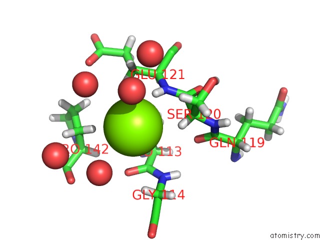
Mono view
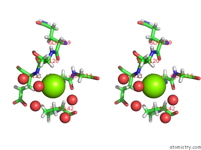
Stereo pair view
|
A full contact list of Magnesium with other atoms in the Mg binding
site number 2 of Crystal Structure of the Pneumoccocal Exposed Lipoprotein Thioredoxin SP_0659 (ETRX1) From Streptococcus Pneumoniae Strain TIGR4 within 5.0Å range:
probe
|
atom
|
residue
|
distance (Å)
|
B
|
Occ
|
A:Mg206
b:24.0
occ:1.00
|
H
|
A:GLU121
|
2.5
|
14.5
|
1.0
|
HA
|
A:SER120
|
2.9
|
14.1
|
1.0
|
HB2
|
A:PRO113
|
3.1
|
14.9
|
1.0
|
O
|
A:HOH465
|
3.2
|
37.4
|
1.0
|
N
|
A:GLU121
|
3.3
|
12.1
|
1.0
|
O
|
A:HOH348
|
3.3
|
23.3
|
1.0
|
HB3
|
A:PRO142
|
3.3
|
17.7
|
1.0
|
HB3
|
A:PRO113
|
3.5
|
14.9
|
1.0
|
HB2
|
A:SER120
|
3.5
|
16.6
|
1.0
|
HB2
|
A:GLU121
|
3.6
|
18.6
|
1.0
|
CA
|
A:SER120
|
3.7
|
11.7
|
1.0
|
CB
|
A:PRO113
|
3.7
|
12.4
|
1.0
|
O
|
A:HOH352
|
3.7
|
27.1
|
1.0
|
HA3
|
A:GLY114
|
3.9
|
15.8
|
1.0
|
C
|
A:SER120
|
3.9
|
12.0
|
1.0
|
C
|
A:PRO113
|
4.0
|
12.7
|
1.0
|
O
|
A:PRO113
|
4.0
|
12.8
|
1.0
|
HB3
|
A:GLU121
|
4.0
|
18.6
|
1.0
|
CB
|
A:SER120
|
4.1
|
13.8
|
1.0
|
CB
|
A:GLU121
|
4.1
|
15.5
|
1.0
|
N
|
A:GLY114
|
4.2
|
11.9
|
1.0
|
CA
|
A:GLU121
|
4.2
|
12.7
|
1.0
|
CB
|
A:PRO142
|
4.3
|
14.7
|
1.0
|
CA
|
A:PRO113
|
4.4
|
10.6
|
1.0
|
CA
|
A:GLY114
|
4.5
|
13.2
|
1.0
|
O
|
A:HOH336
|
4.5
|
19.1
|
1.0
|
HB3
|
A:SER120
|
4.5
|
16.6
|
1.0
|
H
|
A:GLY114
|
4.6
|
14.2
|
1.0
|
HB2
|
A:PRO142
|
4.6
|
17.7
|
1.0
|
HA
|
A:GLU121
|
4.6
|
15.3
|
1.0
|
HA2
|
A:GLY114
|
4.6
|
15.8
|
1.0
|
HA
|
A:PRO142
|
4.7
|
15.5
|
1.0
|
O
|
A:HOH444
|
4.8
|
36.3
|
1.0
|
O
|
A:GLN119
|
4.8
|
11.2
|
1.0
|
HG3
|
A:PRO142
|
4.8
|
25.1
|
1.0
|
N
|
A:SER120
|
4.9
|
10.6
|
1.0
|
HA
|
A:PRO113
|
5.0
|
12.7
|
1.0
|
HG2
|
A:PRO113
|
5.0
|
13.7
|
1.0
|
CG
|
A:PRO113
|
5.0
|
11.4
|
1.0
|
|
Magnesium binding site 3 out
of 9 in 4hqs
Go back to
Magnesium Binding Sites List in 4hqs
Magnesium binding site 3 out
of 9 in the Crystal Structure of the Pneumoccocal Exposed Lipoprotein Thioredoxin SP_0659 (ETRX1) From Streptococcus Pneumoniae Strain TIGR4
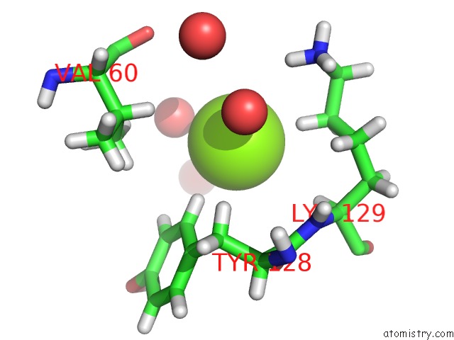
Mono view
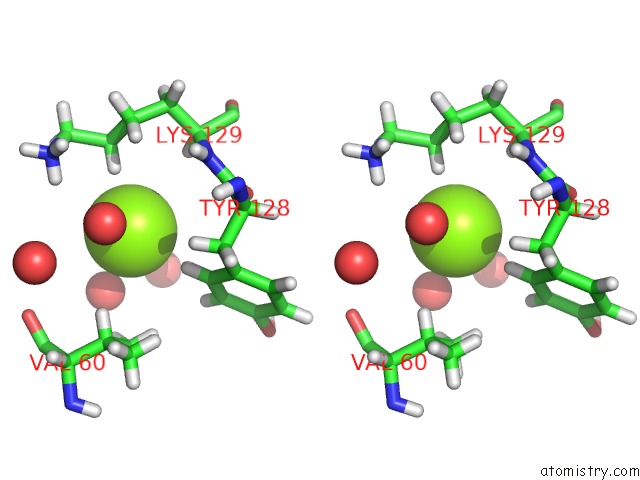
Stereo pair view
|
A full contact list of Magnesium with other atoms in the Mg binding
site number 3 of Crystal Structure of the Pneumoccocal Exposed Lipoprotein Thioredoxin SP_0659 (ETRX1) From Streptococcus Pneumoniae Strain TIGR4 within 5.0Å range:
probe
|
atom
|
residue
|
distance (Å)
|
B
|
Occ
|
A:Mg207
b:23.4
occ:1.00
|
HD3
|
A:LYS129
|
2.6
|
33.3
|
1.0
|
O
|
A:HOH479
|
2.9
|
22.0
|
1.0
|
HD2
|
A:TYR128
|
2.9
|
11.1
|
1.0
|
O
|
A:HOH393
|
3.0
|
24.3
|
1.0
|
CD
|
A:LYS129
|
3.5
|
27.7
|
1.0
|
HD2
|
A:LYS129
|
3.6
|
33.3
|
1.0
|
HA
|
A:LYS129
|
3.7
|
11.9
|
1.0
|
CD2
|
A:TYR128
|
3.7
|
9.2
|
1.0
|
HE2
|
A:TYR128
|
3.8
|
11.2
|
1.0
|
HZ3
|
A:LYS129
|
4.0
|
31.9
|
1.0
|
HB
|
A:VAL60
|
4.0
|
15.7
|
1.0
|
HG23
|
A:VAL60
|
4.0
|
17.5
|
1.0
|
HG21
|
A:VAL60
|
4.1
|
17.5
|
1.0
|
CE2
|
A:TYR128
|
4.2
|
9.4
|
1.0
|
HE3
|
A:LYS129
|
4.2
|
34.4
|
1.0
|
O
|
A:HOH438
|
4.3
|
29.2
|
1.0
|
CE
|
A:LYS129
|
4.3
|
28.7
|
1.0
|
HB3
|
A:TYR128
|
4.4
|
11.6
|
1.0
|
HG2
|
A:LYS129
|
4.4
|
18.7
|
1.0
|
CG2
|
A:VAL60
|
4.4
|
14.6
|
1.0
|
CA
|
A:LYS129
|
4.5
|
9.9
|
1.0
|
CG
|
A:LYS129
|
4.5
|
15.6
|
1.0
|
NZ
|
A:LYS129
|
4.6
|
26.6
|
1.0
|
N
|
A:LYS129
|
4.6
|
10.4
|
1.0
|
O
|
A:TYR128
|
4.6
|
10.9
|
1.0
|
C
|
A:TYR128
|
4.7
|
9.4
|
1.0
|
CB
|
A:VAL60
|
4.7
|
13.1
|
1.0
|
O
|
A:HOH362
|
4.8
|
21.6
|
1.0
|
CG
|
A:TYR128
|
4.8
|
9.5
|
1.0
|
H
|
A:LYS129
|
4.9
|
12.4
|
1.0
|
HZ1
|
A:LYS129
|
4.9
|
31.9
|
1.0
|
O
|
A:VAL60
|
5.0
|
15.5
|
1.0
|
|
Magnesium binding site 4 out
of 9 in 4hqs
Go back to
Magnesium Binding Sites List in 4hqs
Magnesium binding site 4 out
of 9 in the Crystal Structure of the Pneumoccocal Exposed Lipoprotein Thioredoxin SP_0659 (ETRX1) From Streptococcus Pneumoniae Strain TIGR4
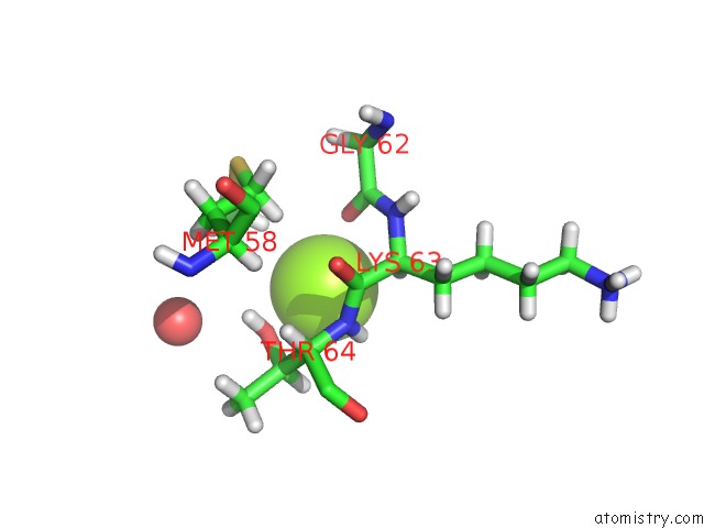
Mono view
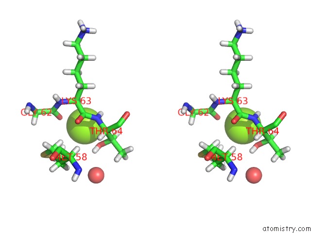
Stereo pair view
|
A full contact list of Magnesium with other atoms in the Mg binding
site number 4 of Crystal Structure of the Pneumoccocal Exposed Lipoprotein Thioredoxin SP_0659 (ETRX1) From Streptococcus Pneumoniae Strain TIGR4 within 5.0Å range:
probe
|
atom
|
residue
|
distance (Å)
|
B
|
Occ
|
A:Mg208
b:18.6
occ:1.00
|
OG1
|
A:THR64
|
2.7
|
12.8
|
1.0
|
O
|
A:GLY62
|
2.7
|
11.2
|
1.0
|
HE1
|
A:MET58
|
2.9
|
18.6
|
1.0
|
H
|
A:THR64
|
3.0
|
12.8
|
1.0
|
HA
|
A:LYS63
|
3.1
|
12.8
|
1.0
|
HG1
|
A:THR64
|
3.2
|
15.3
|
1.0
|
N
|
A:THR64
|
3.3
|
10.7
|
1.0
|
HB
|
A:THR64
|
3.5
|
15.2
|
1.0
|
CB
|
A:THR64
|
3.6
|
12.6
|
1.0
|
C
|
A:LYS63
|
3.7
|
12.1
|
1.0
|
CA
|
A:LYS63
|
3.8
|
10.7
|
1.0
|
CE
|
A:MET58
|
3.8
|
15.5
|
1.0
|
C
|
A:GLY62
|
3.9
|
11.8
|
1.0
|
CA
|
A:THR64
|
4.0
|
12.0
|
1.0
|
HE2
|
A:MET58
|
4.1
|
18.6
|
1.0
|
HG3
|
A:MET58
|
4.1
|
13.8
|
1.0
|
HE3
|
A:MET58
|
4.2
|
18.6
|
1.0
|
N
|
A:LYS63
|
4.4
|
11.0
|
1.0
|
HA
|
A:THR64
|
4.4
|
14.4
|
1.0
|
O
|
A:LYS63
|
4.6
|
12.6
|
1.0
|
O
|
A:HOH333
|
4.8
|
16.4
|
1.0
|
CG2
|
A:THR64
|
4.9
|
13.6
|
1.0
|
CG
|
A:MET58
|
5.0
|
11.5
|
1.0
|
|
Magnesium binding site 5 out
of 9 in 4hqs
Go back to
Magnesium Binding Sites List in 4hqs
Magnesium binding site 5 out
of 9 in the Crystal Structure of the Pneumoccocal Exposed Lipoprotein Thioredoxin SP_0659 (ETRX1) From Streptococcus Pneumoniae Strain TIGR4
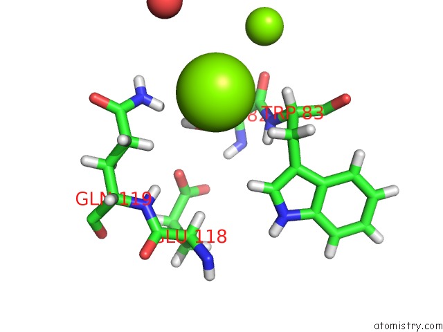
Mono view
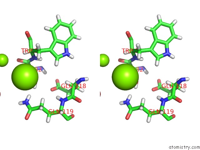
Stereo pair view
|
A full contact list of Magnesium with other atoms in the Mg binding
site number 5 of Crystal Structure of the Pneumoccocal Exposed Lipoprotein Thioredoxin SP_0659 (ETRX1) From Streptococcus Pneumoniae Strain TIGR4 within 5.0Å range:
probe
|
atom
|
residue
|
distance (Å)
|
B
|
Occ
|
A:Mg209
b:21.8
occ:1.00
|
HG
|
A:SER82
|
2.8
|
11.3
|
1.0
|
OG
|
A:SER82
|
2.8
|
9.4
|
1.0
|
HB3
|
A:TRP83
|
3.1
|
11.9
|
1.0
|
HE22
|
A:GLN119
|
3.1
|
17.8
|
1.0
|
HA
|
A:TRP83
|
3.3
|
11.0
|
1.0
|
HB2
|
A:SER82
|
3.4
|
11.6
|
1.0
|
N
|
A:TRP83
|
3.6
|
10.1
|
1.0
|
NE2
|
A:GLN119
|
3.6
|
14.8
|
1.0
|
CB
|
A:SER82
|
3.6
|
9.7
|
1.0
|
C
|
A:SER82
|
3.7
|
9.5
|
1.0
|
CA
|
A:TRP83
|
3.7
|
9.1
|
1.0
|
HG3
|
A:GLN119
|
3.8
|
13.3
|
1.0
|
CB
|
A:TRP83
|
3.8
|
10.0
|
1.0
|
O
|
A:SER82
|
3.9
|
10.7
|
1.0
|
H
|
A:TRP83
|
3.9
|
12.1
|
1.0
|
HE21
|
A:GLN119
|
4.0
|
17.8
|
1.0
|
MG
|
A:MG213
|
4.0
|
26.8
|
1.0
|
CA
|
A:SER82
|
4.3
|
9.1
|
1.0
|
CD
|
A:GLN119
|
4.4
|
12.1
|
1.0
|
HD1
|
A:TRP83
|
4.4
|
12.6
|
1.0
|
HB2
|
A:TRP83
|
4.4
|
11.9
|
1.0
|
CG
|
A:GLN119
|
4.4
|
11.1
|
1.0
|
HB3
|
A:SER82
|
4.4
|
11.6
|
1.0
|
O
|
A:HOH413
|
4.5
|
30.6
|
1.0
|
HG2
|
A:GLN119
|
4.5
|
13.3
|
1.0
|
CG
|
A:TRP83
|
4.8
|
10.8
|
1.0
|
OE1
|
A:GLU118
|
4.9
|
10.3
|
1.0
|
CD1
|
A:TRP83
|
4.9
|
10.5
|
1.0
|
|
Magnesium binding site 6 out
of 9 in 4hqs
Go back to
Magnesium Binding Sites List in 4hqs
Magnesium binding site 6 out
of 9 in the Crystal Structure of the Pneumoccocal Exposed Lipoprotein Thioredoxin SP_0659 (ETRX1) From Streptococcus Pneumoniae Strain TIGR4
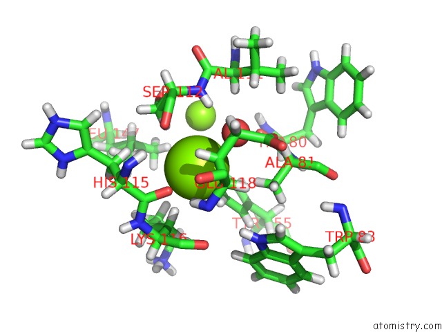
Mono view
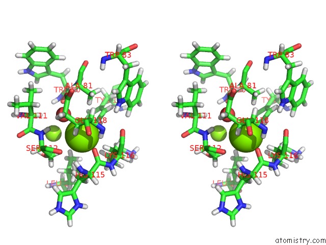
Stereo pair view
|
A full contact list of Magnesium with other atoms in the Mg binding
site number 6 of Crystal Structure of the Pneumoccocal Exposed Lipoprotein Thioredoxin SP_0659 (ETRX1) From Streptococcus Pneumoniae Strain TIGR4 within 5.0Å range:
probe
|
atom
|
residue
|
distance (Å)
|
B
|
Occ
|
A:Mg210
b:20.6
occ:1.00
|
OH
|
A:TYR155
|
2.5
|
10.7
|
1.0
|
HB2
|
A:LYS116
|
2.6
|
16.0
|
1.0
|
HB2
|
A:GLU118
|
2.8
|
10.6
|
1.0
|
O
|
A:HIS115
|
2.8
|
10.6
|
1.0
|
O
|
A:HOH328
|
2.8
|
9.4
|
1.0
|
HE2
|
A:TYR155
|
2.8
|
12.8
|
1.0
|
HB2
|
A:SER112
|
2.8
|
11.4
|
1.0
|
HH
|
A:TYR155
|
3.1
|
12.8
|
1.0
|
CZ
|
A:TYR155
|
3.3
|
8.3
|
1.0
|
CE2
|
A:TYR155
|
3.4
|
10.7
|
1.0
|
HB3
|
A:ALA81
|
3.4
|
11.0
|
1.0
|
H
|
A:SER112
|
3.5
|
10.1
|
1.0
|
O
|
A:SER112
|
3.5
|
10.9
|
1.0
|
CB
|
A:GLU118
|
3.6
|
8.8
|
1.0
|
CB
|
A:LYS116
|
3.6
|
13.3
|
1.0
|
HB3
|
A:GLU118
|
3.6
|
10.6
|
1.0
|
C
|
A:HIS115
|
3.7
|
14.0
|
1.0
|
CB
|
A:SER112
|
3.8
|
9.5
|
1.0
|
HD11
|
A:LEU147
|
3.8
|
17.2
|
1.0
|
HB2
|
A:HIS115
|
3.9
|
15.2
|
1.0
|
H
|
A:GLU118
|
3.9
|
13.3
|
1.0
|
HB3
|
A:LYS116
|
4.0
|
16.0
|
1.0
|
HD3
|
A:LYS116
|
4.0
|
25.6
|
1.0
|
N
|
A:SER112
|
4.1
|
8.4
|
1.0
|
HG2
|
A:GLU118
|
4.1
|
11.4
|
1.0
|
C
|
A:SER112
|
4.2
|
10.4
|
1.0
|
HB2
|
A:ALA81
|
4.2
|
11.0
|
1.0
|
O
|
A:LYS116
|
4.2
|
13.8
|
1.0
|
CB
|
A:ALA81
|
4.2
|
9.2
|
1.0
|
CA
|
A:SER112
|
4.2
|
9.8
|
1.0
|
MG
|
A:MG205
|
4.2
|
17.1
|
1.0
|
CA
|
A:LYS116
|
4.3
|
12.8
|
1.0
|
HB3
|
A:SER112
|
4.3
|
11.4
|
1.0
|
N
|
A:LYS116
|
4.3
|
12.9
|
1.0
|
C
|
A:LYS116
|
4.4
|
11.7
|
1.0
|
HG3
|
A:LYS116
|
4.4
|
19.1
|
1.0
|
H
|
A:HIS115
|
4.4
|
13.8
|
1.0
|
CG
|
A:GLU118
|
4.4
|
9.5
|
1.0
|
HE1
|
A:TRP83
|
4.5
|
12.9
|
1.0
|
CG
|
A:LYS116
|
4.5
|
15.9
|
1.0
|
N
|
A:GLU118
|
4.5
|
11.1
|
1.0
|
CB
|
A:HIS115
|
4.5
|
12.7
|
1.0
|
OG
|
A:SER112
|
4.6
|
10.6
|
1.0
|
O
|
A:TRP80
|
4.6
|
7.7
|
1.0
|
HB3
|
A:HIS115
|
4.6
|
15.2
|
1.0
|
CA
|
A:HIS115
|
4.6
|
11.8
|
1.0
|
CA
|
A:GLU118
|
4.6
|
11.0
|
1.0
|
CE1
|
A:TYR155
|
4.6
|
9.7
|
1.0
|
HD22
|
A:LEU147
|
4.7
|
18.3
|
1.0
|
CD2
|
A:TYR155
|
4.7
|
11.4
|
1.0
|
CD1
|
A:LEU147
|
4.7
|
14.4
|
1.0
|
OE2
|
A:GLU118
|
4.7
|
9.2
|
1.0
|
CD
|
A:LYS116
|
4.7
|
21.3
|
1.0
|
HA
|
A:ALA81
|
4.7
|
10.2
|
1.0
|
HD13
|
A:LEU147
|
4.8
|
17.2
|
1.0
|
HB1
|
A:ALA81
|
4.9
|
11.0
|
1.0
|
HA
|
A:GLU118
|
4.9
|
13.2
|
1.0
|
N
|
A:HIS115
|
4.9
|
11.5
|
1.0
|
HA
|
A:VAL111
|
4.9
|
9.3
|
1.0
|
HG
|
A:SER112
|
5.0
|
12.7
|
1.0
|
HE1
|
A:TYR155
|
5.0
|
11.7
|
1.0
|
CD
|
A:GLU118
|
5.0
|
8.4
|
1.0
|
|
Magnesium binding site 7 out
of 9 in 4hqs
Go back to
Magnesium Binding Sites List in 4hqs
Magnesium binding site 7 out
of 9 in the Crystal Structure of the Pneumoccocal Exposed Lipoprotein Thioredoxin SP_0659 (ETRX1) From Streptococcus Pneumoniae Strain TIGR4
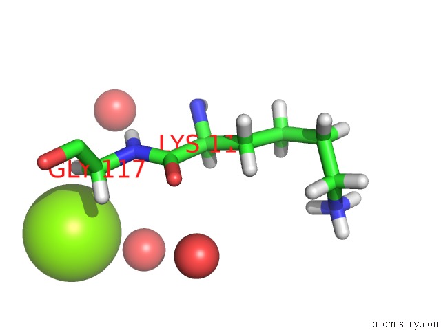
Mono view
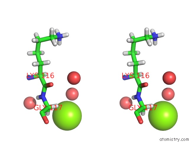
Stereo pair view
|
A full contact list of Magnesium with other atoms in the Mg binding
site number 7 of Crystal Structure of the Pneumoccocal Exposed Lipoprotein Thioredoxin SP_0659 (ETRX1) From Streptococcus Pneumoniae Strain TIGR4 within 5.0Å range:
probe
|
atom
|
residue
|
distance (Å)
|
B
|
Occ
|
A:Mg211
b:29.8
occ:1.00
|
O
|
A:HOH519
|
2.6
|
35.5
|
1.0
|
HA2
|
A:GLY117
|
2.9
|
15.1
|
1.0
|
HA3
|
A:GLY117
|
3.2
|
15.1
|
1.0
|
CA
|
A:GLY117
|
3.5
|
12.6
|
1.0
|
N
|
A:GLY117
|
4.1
|
13.1
|
1.0
|
H
|
A:GLY117
|
4.2
|
15.7
|
1.0
|
O
|
A:HOH368
|
4.6
|
21.0
|
1.0
|
C
|
A:GLY117
|
4.7
|
13.1
|
1.0
|
O
|
A:HOH354
|
4.8
|
24.0
|
1.0
|
C
|
A:LYS116
|
4.9
|
11.7
|
1.0
|
O
|
A:GLY117
|
5.0
|
11.7
|
1.0
|
|
Magnesium binding site 8 out
of 9 in 4hqs
Go back to
Magnesium Binding Sites List in 4hqs
Magnesium binding site 8 out
of 9 in the Crystal Structure of the Pneumoccocal Exposed Lipoprotein Thioredoxin SP_0659 (ETRX1) From Streptococcus Pneumoniae Strain TIGR4
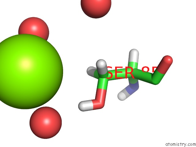
Mono view
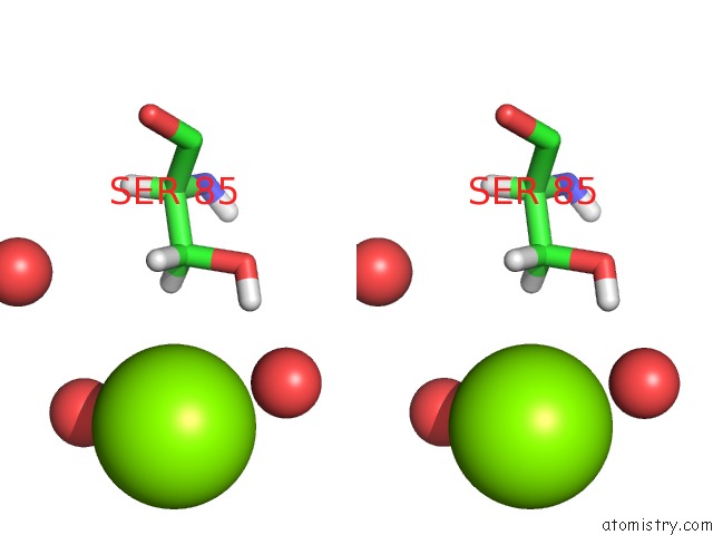
Stereo pair view
|
A full contact list of Magnesium with other atoms in the Mg binding
site number 8 of Crystal Structure of the Pneumoccocal Exposed Lipoprotein Thioredoxin SP_0659 (ETRX1) From Streptococcus Pneumoniae Strain TIGR4 within 5.0Å range:
probe
|
atom
|
residue
|
distance (Å)
|
B
|
Occ
|
A:Mg212
b:33.7
occ:1.00
|
O
|
A:HOH497
|
2.5
|
37.6
|
1.0
|
O
|
A:HOH436
|
2.6
|
30.6
|
1.0
|
HG
|
A:SER85
|
3.2
|
29.3
|
1.0
|
HB2
|
A:SER85
|
3.6
|
22.7
|
1.0
|
HB3
|
A:SER85
|
3.6
|
22.7
|
1.0
|
CB
|
A:SER85
|
3.9
|
18.9
|
1.0
|
OG
|
A:SER85
|
3.9
|
24.4
|
1.0
|
O
|
A:HOH458
|
3.9
|
37.0
|
1.0
|
O
|
A:HOH460
|
4.9
|
27.8
|
1.0
|
|
Magnesium binding site 9 out
of 9 in 4hqs
Go back to
Magnesium Binding Sites List in 4hqs
Magnesium binding site 9 out
of 9 in the Crystal Structure of the Pneumoccocal Exposed Lipoprotein Thioredoxin SP_0659 (ETRX1) From Streptococcus Pneumoniae Strain TIGR4
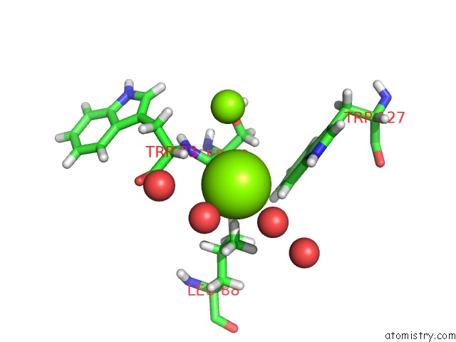
Mono view
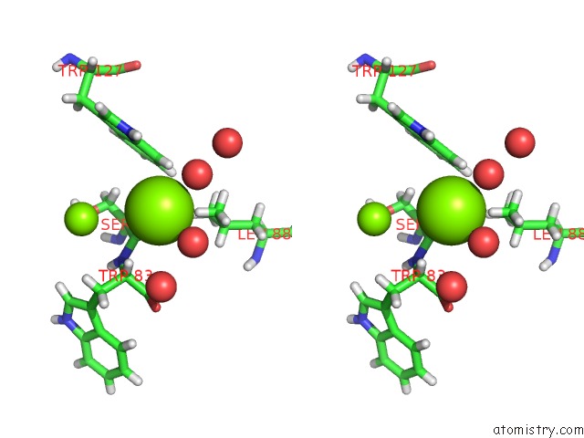
Stereo pair view
|
A full contact list of Magnesium with other atoms in the Mg binding
site number 9 of Crystal Structure of the Pneumoccocal Exposed Lipoprotein Thioredoxin SP_0659 (ETRX1) From Streptococcus Pneumoniae Strain TIGR4 within 5.0Å range:
probe
|
atom
|
residue
|
distance (Å)
|
B
|
Occ
|
A:Mg213
b:26.8
occ:1.00
|
O
|
A:SER82
|
2.7
|
10.7
|
1.0
|
O
|
A:HOH404
|
2.7
|
34.7
|
1.0
|
O
|
A:HOH521
|
2.7
|
33.5
|
1.0
|
HD12
|
A:LEU88
|
2.9
|
17.4
|
1.0
|
HA
|
A:TRP83
|
3.2
|
11.0
|
1.0
|
HZ2
|
A:TRP127
|
3.4
|
13.1
|
1.0
|
CD1
|
A:LEU88
|
3.7
|
14.5
|
1.0
|
HD13
|
A:LEU88
|
3.7
|
17.4
|
1.0
|
C
|
A:SER82
|
3.7
|
9.5
|
1.0
|
HE1
|
A:TRP127
|
3.9
|
14.8
|
1.0
|
HD11
|
A:LEU88
|
4.0
|
17.4
|
1.0
|
MG
|
A:MG209
|
4.0
|
21.8
|
1.0
|
CZ2
|
A:TRP127
|
4.1
|
10.9
|
1.0
|
O
|
A:HOH359
|
4.1
|
23.6
|
1.0
|
CA
|
A:TRP83
|
4.1
|
9.1
|
1.0
|
HB2
|
A:SER82
|
4.3
|
11.6
|
1.0
|
N
|
A:TRP83
|
4.3
|
10.1
|
1.0
|
NE1
|
A:TRP127
|
4.4
|
12.3
|
1.0
|
CE2
|
A:TRP127
|
4.5
|
10.6
|
1.0
|
C
|
A:TRP83
|
4.6
|
11.8
|
1.0
|
O
|
A:HOH394
|
4.8
|
24.6
|
1.0
|
HD21
|
A:LEU88
|
4.8
|
18.4
|
1.0
|
CA
|
A:SER82
|
4.8
|
9.1
|
1.0
|
HG
|
A:LEU88
|
4.9
|
15.3
|
1.0
|
CG
|
A:LEU88
|
4.9
|
12.7
|
1.0
|
HA
|
A:SER82
|
4.9
|
10.9
|
1.0
|
O
|
A:TRP83
|
4.9
|
13.1
|
1.0
|
CB
|
A:SER82
|
5.0
|
9.7
|
1.0
|
|
Reference:
M.Saleh,
S.G.Bartual,
M.R.Abdullah,
I.Jensch,
T.M.Asmat,
L.Petruschka,
T.Pribyl,
M.Gellert,
C.H.Lillig,
H.Antelmann,
J.A.Hermoso,
S.Hammerschmidt.
Molecular Architecture of Streptococcus Pneumoniae Surface Thioredoxin-Fold Lipoproteins Crucial For Extracellular Oxidative Stress Resistance and Maintenance of Virulence. Embo Mol Med V. 5 1852 2013.
ISSN: ISSN 1757-4676
PubMed: 24136784
DOI: 10.1002/EMMM.201202435
Page generated: Fri Aug 16 16:23:08 2024
|