Magnesium »
PDB 4hmy-4hyv »
4hqx »
Magnesium in PDB 4hqx: Crystal Structure of Human Pdgf-Bb in Complex with A Modified Nucleotide Aptamer (Somamer SL4)
Protein crystallography data
The structure of Crystal Structure of Human Pdgf-Bb in Complex with A Modified Nucleotide Aptamer (Somamer SL4), PDB code: 4hqx
was solved by
D.R.Davies,
T.E.Edwards,
N.Janjic,
A.D.Gelinas,
C.Zhang,
T.C.Jarvis,
with X-Ray Crystallography technique. A brief refinement statistics is given in the table below:
| Resolution Low / High (Å) | 19.68 / 2.30 |
| Space group | P 41 21 2 |
| Cell size a, b, c (Å), α, β, γ (°) | 59.430, 59.430, 168.160, 90.00, 90.00, 90.00 |
| R / Rfree (%) | 23.9 / 27.9 |
Other elements in 4hqx:
The structure of Crystal Structure of Human Pdgf-Bb in Complex with A Modified Nucleotide Aptamer (Somamer SL4) also contains other interesting chemical elements:
| Sodium | (Na) | 1 atom |
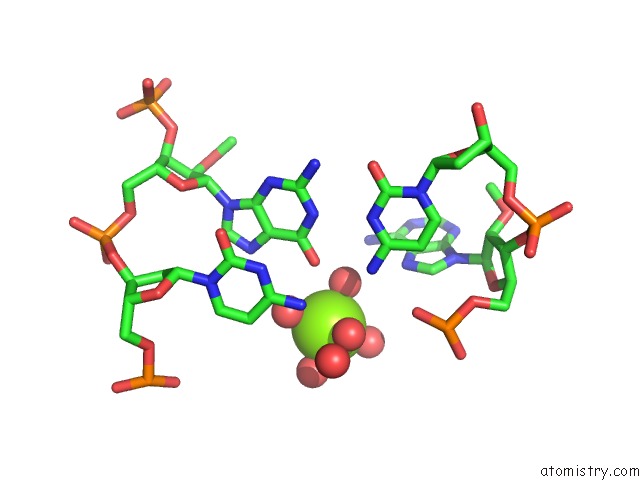
Magnesium Binding Sites:
The binding sites of Magnesium atom in the Crystal Structure of Human Pdgf-Bb in Complex with A Modified Nucleotide Aptamer (Somamer SL4)
(pdb code 4hqx). This binding sites where shown within
5.0 Angstroms radius around Magnesium atom.
In total only one binding site of Magnesium was determined in the Crystal Structure of Human Pdgf-Bb in Complex with A Modified Nucleotide Aptamer (Somamer SL4), PDB code: 4hqx:
In total only one binding site of Magnesium was determined in the Crystal Structure of Human Pdgf-Bb in Complex with A Modified Nucleotide Aptamer (Somamer SL4), PDB code: 4hqx:
Magnesium binding site 1 out of 1 in 4hqx
Go back to
Magnesium binding site 1 out
of 1 in the Crystal Structure of Human Pdgf-Bb in Complex with A Modified Nucleotide Aptamer (Somamer SL4)
Mono view
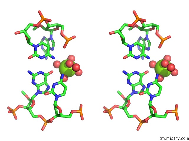
Stereo pair view
Mono view
Stereo pair view
A full contact list of Magnesium with other atoms in the Mg binding
site number 1 of Crystal Structure of Human Pdgf-Bb in Complex with A Modified Nucleotide Aptamer (Somamer SL4) within 5.0Å range:
|
Reference:
D.R.Davies,
A.D.Gelinas,
C.Zhang,
J.C.Rohloff,
J.D.Carter,
D.O'connell,
S.M.Waugh,
S.K.Wolk,
W.S.Mayfield,
A.B.Burgin,
T.E.Edwards,
L.J.Stewart,
L.Gold,
N.Janjic,
T.C.Jarvis.
Unique Motifs and Hydrophobic Interactions Shape the Binding of Modified Dna Ligands to Protein Targets. Proc.Natl.Acad.Sci.Usa V. 109 19971 2012.
ISSN: ISSN 0027-8424
PubMed: 23139410
DOI: 10.1073/PNAS.1213933109
Page generated: Mon Aug 11 13:55:32 2025
ISSN: ISSN 0027-8424
PubMed: 23139410
DOI: 10.1073/PNAS.1213933109
Last articles
Mg in 5E6SMg in 5E75
Mg in 5E6U
Mg in 5E67
Mg in 5E6F
Mg in 5E6R
Mg in 5E54
Mg in 5E63
Mg in 5E5A
Mg in 5E5S