Magnesium »
PDB 4hyw-4iaj »
4i7z »
Magnesium in PDB 4i7z: Crystal Structure of Cytochrome B6F in Dopg, with Disordered Rieske Iron-Sulfur Protein Soluble Domain
Enzymatic activity of Crystal Structure of Cytochrome B6F in Dopg, with Disordered Rieske Iron-Sulfur Protein Soluble Domain
All present enzymatic activity of Crystal Structure of Cytochrome B6F in Dopg, with Disordered Rieske Iron-Sulfur Protein Soluble Domain:
1.10.9.1;
1.10.9.1;
Protein crystallography data
The structure of Crystal Structure of Cytochrome B6F in Dopg, with Disordered Rieske Iron-Sulfur Protein Soluble Domain, PDB code: 4i7z
was solved by
S.S.Hasan,
J.T.Stofleth,
E.Yamashita,
W.A.Cramer,
with X-Ray Crystallography technique. A brief refinement statistics is given in the table below:
| Resolution Low / High (Å) | 48.52 / 2.80 |
| Space group | P 61 2 2 |
| Cell size a, b, c (Å), α, β, γ (°) | 159.451, 159.451, 362.746, 90.00, 90.00, 120.00 |
| R / Rfree (%) | 24.8 / 27.2 |
Other elements in 4i7z:
The structure of Crystal Structure of Cytochrome B6F in Dopg, with Disordered Rieske Iron-Sulfur Protein Soluble Domain also contains other interesting chemical elements:
| Cadmium | (Cd) | 3 atoms |
| Iron | (Fe) | 4 atoms |
Magnesium Binding Sites:
The binding sites of Magnesium atom in the Crystal Structure of Cytochrome B6F in Dopg, with Disordered Rieske Iron-Sulfur Protein Soluble Domain
(pdb code 4i7z). This binding sites where shown within
5.0 Angstroms radius around Magnesium atom.
In total only one binding site of Magnesium was determined in the Crystal Structure of Cytochrome B6F in Dopg, with Disordered Rieske Iron-Sulfur Protein Soluble Domain, PDB code: 4i7z:
In total only one binding site of Magnesium was determined in the Crystal Structure of Cytochrome B6F in Dopg, with Disordered Rieske Iron-Sulfur Protein Soluble Domain, PDB code: 4i7z:
Magnesium binding site 1 out of 1 in 4i7z
Go back to
Magnesium binding site 1 out
of 1 in the Crystal Structure of Cytochrome B6F in Dopg, with Disordered Rieske Iron-Sulfur Protein Soluble Domain
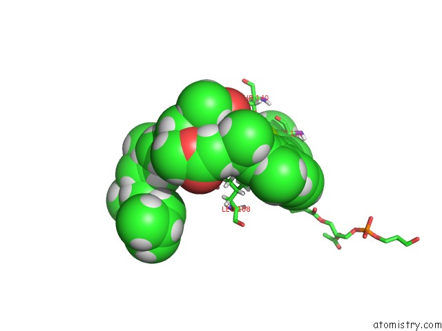
Mono view
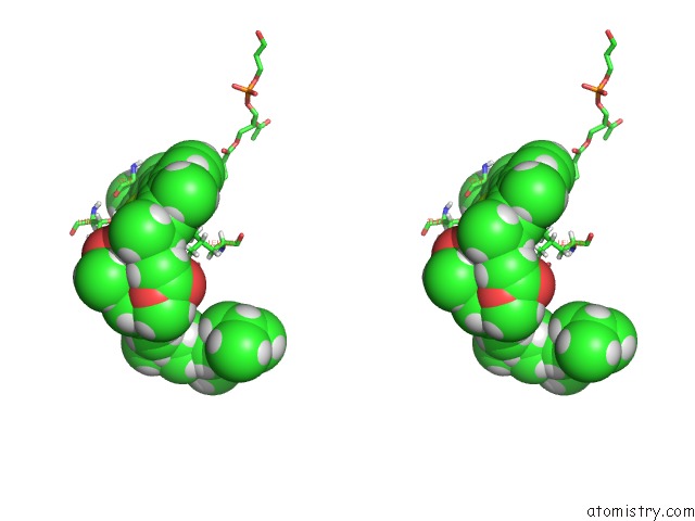
Stereo pair view
Mono view
Stereo pair view
A full contact list of Magnesium with other atoms in the Mg binding
site number 1 of Crystal Structure of Cytochrome B6F in Dopg, with Disordered Rieske Iron-Sulfur Protein Soluble Domain within 5.0Å range:
|
Reference:
S.S.Hasan,
J.T.Stofleth,
E.Yamashita,
W.A.Cramer.
Lipid-Induced Conformational Changes Within the Cytochrome B6F Complex of Oxygenic Photosynthesis. Biochemistry V. 52 2649 2013.
ISSN: ISSN 0006-2960
PubMed: 23514009
DOI: 10.1021/BI301638H
Page generated: Fri Aug 16 16:36:49 2024
ISSN: ISSN 0006-2960
PubMed: 23514009
DOI: 10.1021/BI301638H
Last articles
Zn in 9MJ5Zn in 9HNW
Zn in 9G0L
Zn in 9FNE
Zn in 9DZN
Zn in 9E0I
Zn in 9D32
Zn in 9DAK
Zn in 8ZXC
Zn in 8ZUF