Magnesium »
PDB 4ibd-4iir »
4if4 »
Magnesium in PDB 4if4: Crystal Structure of the Magnesium and Beryllofluoride-Activated Vrar From Staphylococcus Aureus
Protein crystallography data
The structure of Crystal Structure of the Magnesium and Beryllofluoride-Activated Vrar From Staphylococcus Aureus, PDB code: 4if4
was solved by
P.G.Leonard,
A.M.Stock,
with X-Ray Crystallography technique. A brief refinement statistics is given in the table below:
| Resolution Low / High (Å) | 35.98 / 2.35 |
| Space group | H 3 |
| Cell size a, b, c (Å), α, β, γ (°) | 110.603, 110.603, 284.292, 90.00, 90.00, 120.00 |
| R / Rfree (%) | 18.1 / 22.6 |
Other elements in 4if4:
The structure of Crystal Structure of the Magnesium and Beryllofluoride-Activated Vrar From Staphylococcus Aureus also contains other interesting chemical elements:
| Fluorine | (F) | 12 atoms |
Magnesium Binding Sites:
The binding sites of Magnesium atom in the Crystal Structure of the Magnesium and Beryllofluoride-Activated Vrar From Staphylococcus Aureus
(pdb code 4if4). This binding sites where shown within
5.0 Angstroms radius around Magnesium atom.
In total 4 binding sites of Magnesium where determined in the Crystal Structure of the Magnesium and Beryllofluoride-Activated Vrar From Staphylococcus Aureus, PDB code: 4if4:
Jump to Magnesium binding site number: 1; 2; 3; 4;
In total 4 binding sites of Magnesium where determined in the Crystal Structure of the Magnesium and Beryllofluoride-Activated Vrar From Staphylococcus Aureus, PDB code: 4if4:
Jump to Magnesium binding site number: 1; 2; 3; 4;
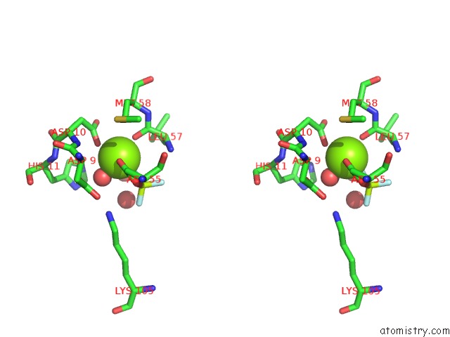
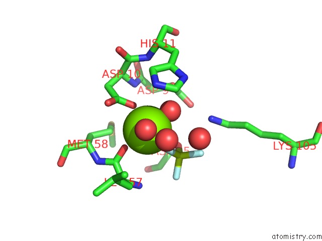
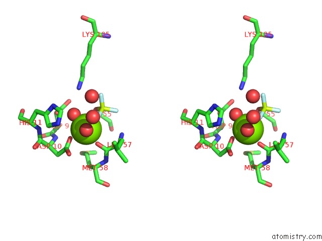
Magnesium binding site 1 out of 4 in 4if4
Go back to
Magnesium binding site 1 out
of 4 in the Crystal Structure of the Magnesium and Beryllofluoride-Activated Vrar From Staphylococcus Aureus
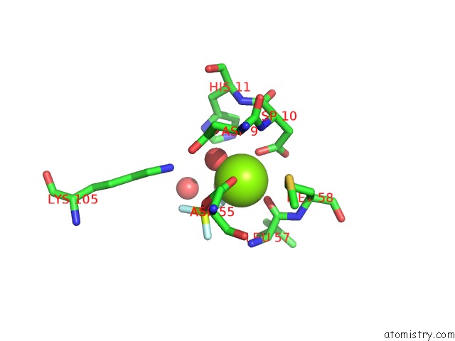
Mono view
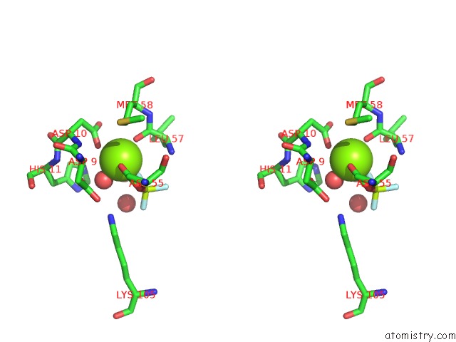
Stereo pair view
Mono view
Stereo pair view
A full contact list of Magnesium with other atoms in the Mg binding
site number 1 of Crystal Structure of the Magnesium and Beryllofluoride-Activated Vrar From Staphylococcus Aureus within 5.0Å range:
|
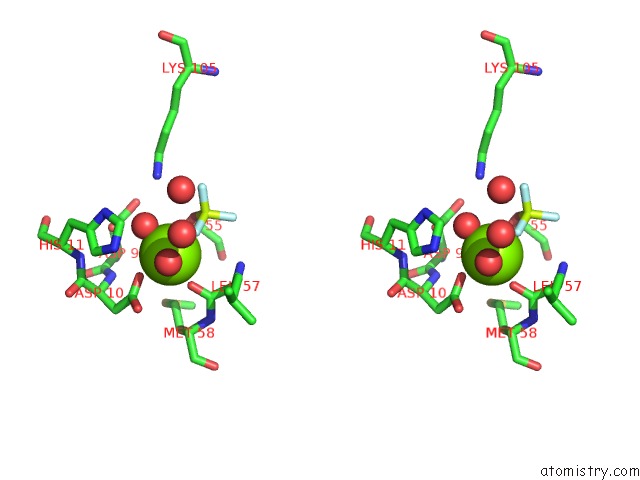
Magnesium binding site 2 out of 4 in 4if4
Go back to
Magnesium binding site 2 out
of 4 in the Crystal Structure of the Magnesium and Beryllofluoride-Activated Vrar From Staphylococcus Aureus
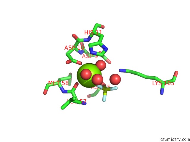
Mono view
Stereo pair view
Mono view
Stereo pair view
A full contact list of Magnesium with other atoms in the Mg binding
site number 2 of Crystal Structure of the Magnesium and Beryllofluoride-Activated Vrar From Staphylococcus Aureus within 5.0Å range:
|
Magnesium binding site 3 out of 4 in 4if4
Go back to
Magnesium binding site 3 out
of 4 in the Crystal Structure of the Magnesium and Beryllofluoride-Activated Vrar From Staphylococcus Aureus

Mono view
Stereo pair view
Mono view
Stereo pair view
A full contact list of Magnesium with other atoms in the Mg binding
site number 3 of Crystal Structure of the Magnesium and Beryllofluoride-Activated Vrar From Staphylococcus Aureus within 5.0Å range:
|
Magnesium binding site 4 out of 4 in 4if4
Go back to
Magnesium binding site 4 out
of 4 in the Crystal Structure of the Magnesium and Beryllofluoride-Activated Vrar From Staphylococcus Aureus
Mono view
Stereo pair view
Mono view
Stereo pair view
A full contact list of Magnesium with other atoms in the Mg binding
site number 4 of Crystal Structure of the Magnesium and Beryllofluoride-Activated Vrar From Staphylococcus Aureus within 5.0Å range:
|
Reference:
P.G.Leonard,
D.Golemi-Kotra,
A.M.Stock.
Phosphorylation-Dependent Conformational Changes and Domain Rearrangements in Staphylococcus Aureus Vrar Activation. Proc.Natl.Acad.Sci.Usa V. 110 8525 2013.
ISSN: ISSN 0027-8424
PubMed: 23650349
DOI: 10.1073/PNAS.1302819110
Page generated: Fri Aug 16 16:40:25 2024
ISSN: ISSN 0027-8424
PubMed: 23650349
DOI: 10.1073/PNAS.1302819110
Last articles
Fe in 2YXOFe in 2YRS
Fe in 2YXC
Fe in 2YNM
Fe in 2YVJ
Fe in 2YP1
Fe in 2YU2
Fe in 2YU1
Fe in 2YQB
Fe in 2YOO