Magnesium »
PDB 4ijm-4ix3 »
4ip2 »
Magnesium in PDB 4ip2: Putative Aromatic Acid Decarboxylase
Protein crystallography data
The structure of Putative Aromatic Acid Decarboxylase, PDB code: 4ip2
was solved by
G.Schneider,
K.Brunner,
A.Izumi,
A.Jacewicz,
with X-Ray Crystallography technique. A brief refinement statistics is given in the table below:
| Resolution Low / High (Å) | 50.30 / 1.95 |
| Space group | P 32 2 1 |
| Cell size a, b, c (Å), α, β, γ (°) | 96.850, 96.850, 301.481, 90.00, 90.00, 120.00 |
| R / Rfree (%) | 19.9 / 24.8 |
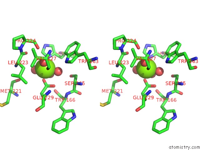
Magnesium Binding Sites:
The binding sites of Magnesium atom in the Putative Aromatic Acid Decarboxylase
(pdb code 4ip2). This binding sites where shown within
5.0 Angstroms radius around Magnesium atom.
In total 3 binding sites of Magnesium where determined in the Putative Aromatic Acid Decarboxylase, PDB code: 4ip2:
Jump to Magnesium binding site number: 1; 2; 3;
In total 3 binding sites of Magnesium where determined in the Putative Aromatic Acid Decarboxylase, PDB code: 4ip2:
Jump to Magnesium binding site number: 1; 2; 3;
Magnesium binding site 1 out of 3 in 4ip2
Go back to
Magnesium binding site 1 out
of 3 in the Putative Aromatic Acid Decarboxylase
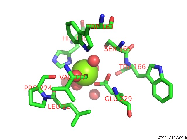
Mono view
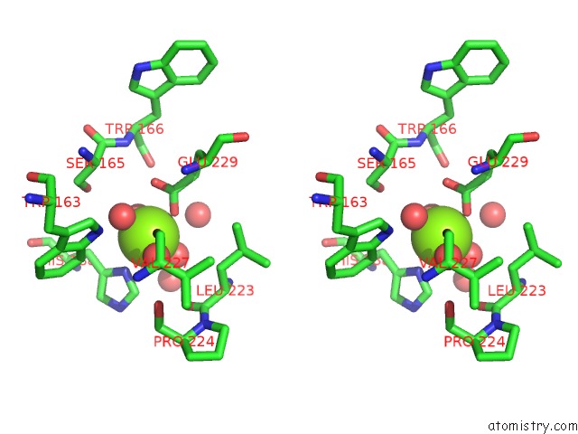
Stereo pair view
Mono view
Stereo pair view
A full contact list of Magnesium with other atoms in the Mg binding
site number 1 of Putative Aromatic Acid Decarboxylase within 5.0Å range:
|
Magnesium binding site 2 out of 3 in 4ip2
Go back to
Magnesium binding site 2 out
of 3 in the Putative Aromatic Acid Decarboxylase
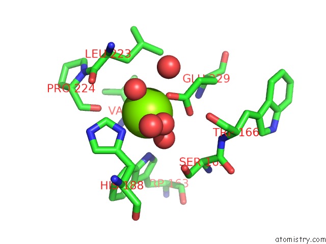
Mono view

Stereo pair view
Mono view
Stereo pair view
A full contact list of Magnesium with other atoms in the Mg binding
site number 2 of Putative Aromatic Acid Decarboxylase within 5.0Å range:
|
Magnesium binding site 3 out of 3 in 4ip2
Go back to
Magnesium binding site 3 out
of 3 in the Putative Aromatic Acid Decarboxylase
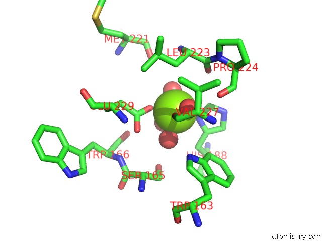
Mono view
Stereo pair view
Mono view
Stereo pair view
A full contact list of Magnesium with other atoms in the Mg binding
site number 3 of Putative Aromatic Acid Decarboxylase within 5.0Å range:
|
Reference:
A.Jacewicz,
A.Izumi,
K.Brunner,
R.Schnell,
G.Schneider.
Structural Insights Into the Ubid Protein Family From the Crystal Structure of PA0254 From Pseudomonas Aeruginosa. Plos One V. 8 63161 2013.
ISSN: ESSN 1932-6203
PubMed: 23671667
DOI: 10.1371/JOURNAL.PONE.0063161
Page generated: Mon Aug 11 14:23:23 2025
ISSN: ESSN 1932-6203
PubMed: 23671667
DOI: 10.1371/JOURNAL.PONE.0063161
Last articles
Mg in 4LF9Mg in 4LF7
Mg in 4LF6
Mg in 4LF4
Mg in 4LF5
Mg in 4LCZ
Mg in 4LF2
Mg in 4LF1
Mg in 4LEM
Mg in 4LCK