Magnesium »
PDB 4ijm-4ix3 »
4itd »
Magnesium in PDB 4itd: Structures of Dna Duplexes Containing O6-Carboxymethylguanine, A Lesion Associated with Gastrointestinal Cancer, Reveal A Mechanism For Inducing Transition Mutation
Protein crystallography data
The structure of Structures of Dna Duplexes Containing O6-Carboxymethylguanine, A Lesion Associated with Gastrointestinal Cancer, Reveal A Mechanism For Inducing Transition Mutation, PDB code: 4itd
was solved by
F.Zhang,
K.Suzuki,
M.Tsunoda,
O.Wilkinson,
C.L.Millington,
D.M.Williams,
E.C.Morishita,
A.Takenaka,
with X-Ray Crystallography technique. A brief refinement statistics is given in the table below:
| Resolution Low / High (Å) | 34.41 / 1.94 |
| Space group | P 21 21 21 |
| Cell size a, b, c (Å), α, β, γ (°) | 25.051, 40.404, 65.667, 90.00, 90.00, 90.00 |
| R / Rfree (%) | 22.6 / 25.4 |
Magnesium Binding Sites:
The binding sites of Magnesium atom in the Structures of Dna Duplexes Containing O6-Carboxymethylguanine, A Lesion Associated with Gastrointestinal Cancer, Reveal A Mechanism For Inducing Transition Mutation
(pdb code 4itd). This binding sites where shown within
5.0 Angstroms radius around Magnesium atom.
In total only one binding site of Magnesium was determined in the Structures of Dna Duplexes Containing O6-Carboxymethylguanine, A Lesion Associated with Gastrointestinal Cancer, Reveal A Mechanism For Inducing Transition Mutation, PDB code: 4itd:
In total only one binding site of Magnesium was determined in the Structures of Dna Duplexes Containing O6-Carboxymethylguanine, A Lesion Associated with Gastrointestinal Cancer, Reveal A Mechanism For Inducing Transition Mutation, PDB code: 4itd:
Magnesium binding site 1 out of 1 in 4itd
Go back to
Magnesium binding site 1 out
of 1 in the Structures of Dna Duplexes Containing O6-Carboxymethylguanine, A Lesion Associated with Gastrointestinal Cancer, Reveal A Mechanism For Inducing Transition Mutation
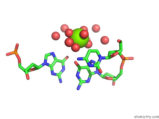
Mono view
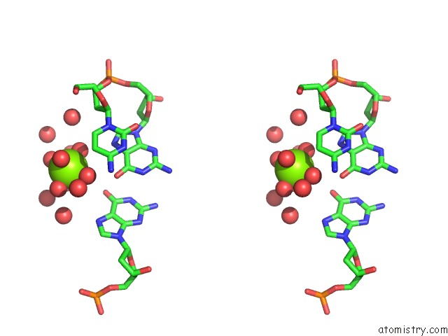
Stereo pair view
Mono view
Stereo pair view
A full contact list of Magnesium with other atoms in the Mg binding
site number 1 of Structures of Dna Duplexes Containing O6-Carboxymethylguanine, A Lesion Associated with Gastrointestinal Cancer, Reveal A Mechanism For Inducing Transition Mutation within 5.0Å range:
|
Reference:
F.Zhang,
M.Tsunoda,
K.Suzuki,
Y.Kikuchi,
O.Wilkinson,
C.L.Millington,
G.P.Margison,
D.M.Williams,
E.Czarina Morishita,
A.Takenaka.
Structures of Dna Duplexes Containing O6-Carboxymethylguanine, A Lesion Associated with Gastrointestinal Cancer, Reveal A Mechanism For Inducing Pyrimidine Transition Mutations Nucleic Acids Res. V. 41 5524 2013.
ISSN: ISSN 0305-1048
PubMed: 23580550
DOI: 10.1093/NAR/GKT198
Page generated: Mon Aug 11 14:26:42 2025
ISSN: ISSN 0305-1048
PubMed: 23580550
DOI: 10.1093/NAR/GKT198
Last articles
Mg in 4JJSMg in 4JJ2
Mg in 4JIW
Mg in 4JIV
Mg in 4JIB
Mg in 4JI4
Mg in 4JI5
Mg in 4JI1
Mg in 4JI0
Mg in 4JI2