Magnesium »
PDB 4ijm-4ix3 »
4ivg »
Magnesium in PDB 4ivg: Crystal Structure of Mitochondrial HSP90 (TRAP1) Ntd-Middle Domain Dimer with Amppnp
Protein crystallography data
The structure of Crystal Structure of Mitochondrial HSP90 (TRAP1) Ntd-Middle Domain Dimer with Amppnp, PDB code: 4ivg
was solved by
J.R.Partridge,
L.A.Lavery,
D.A.Agard,
with X-Ray Crystallography technique. A brief refinement statistics is given in the table below:
| Resolution Low / High (Å) | 29.55 / 1.75 |
| Space group | I 2 2 2 |
| Cell size a, b, c (Å), α, β, γ (°) | 85.146, 94.509, 155.456, 90.00, 90.00, 90.00 |
| R / Rfree (%) | 18.9 / 20.3 |
Magnesium Binding Sites:
The binding sites of Magnesium atom in the Crystal Structure of Mitochondrial HSP90 (TRAP1) Ntd-Middle Domain Dimer with Amppnp
(pdb code 4ivg). This binding sites where shown within
5.0 Angstroms radius around Magnesium atom.
In total only one binding site of Magnesium was determined in the Crystal Structure of Mitochondrial HSP90 (TRAP1) Ntd-Middle Domain Dimer with Amppnp, PDB code: 4ivg:
In total only one binding site of Magnesium was determined in the Crystal Structure of Mitochondrial HSP90 (TRAP1) Ntd-Middle Domain Dimer with Amppnp, PDB code: 4ivg:
Magnesium binding site 1 out of 1 in 4ivg
Go back to
Magnesium binding site 1 out
of 1 in the Crystal Structure of Mitochondrial HSP90 (TRAP1) Ntd-Middle Domain Dimer with Amppnp
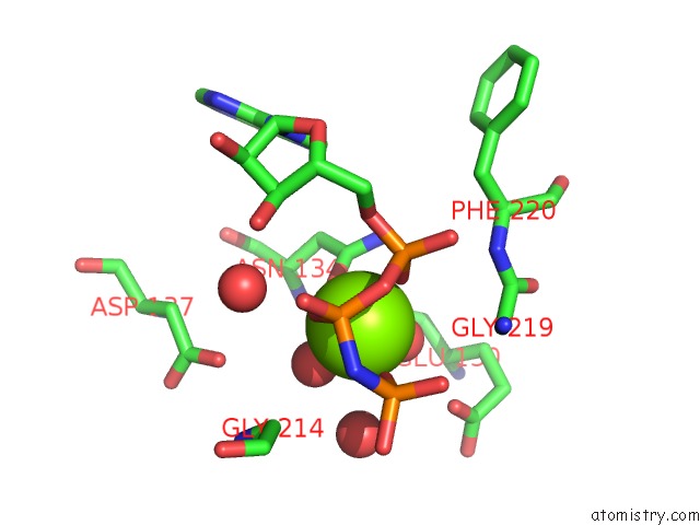
Mono view
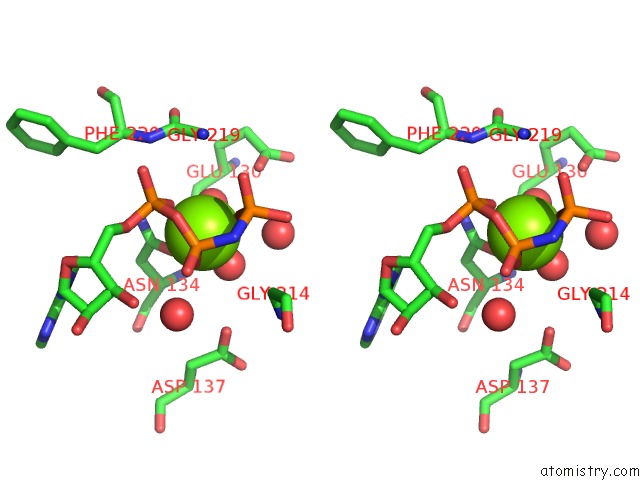
Stereo pair view
Mono view
Stereo pair view
A full contact list of Magnesium with other atoms in the Mg binding
site number 1 of Crystal Structure of Mitochondrial HSP90 (TRAP1) Ntd-Middle Domain Dimer with Amppnp within 5.0Å range:
|
Reference:
L.A.Lavery,
J.R.Partridge,
T.A.Ramelot,
D.Elnatan,
M.A.Kennedy,
D.A.Agard.
Structural Asymmetry in the Closed State of Mitochondrial HSP90 (TRAP1) Supports A Two-Step Atp Hydrolysis Mechanism. Mol.Cell V. 53 330 2014.
ISSN: ISSN 1097-2765
PubMed: 24462206
DOI: 10.1016/J.MOLCEL.2013.12.023
Page generated: Mon Aug 11 14:27:27 2025
ISSN: ISSN 1097-2765
PubMed: 24462206
DOI: 10.1016/J.MOLCEL.2013.12.023
Last articles
Mg in 4LF9Mg in 4LF7
Mg in 4LF6
Mg in 4LF4
Mg in 4LF5
Mg in 4LCZ
Mg in 4LF2
Mg in 4LF1
Mg in 4LEM
Mg in 4LCK