Magnesium »
PDB 4ix3-4j99 »
4j43 »
Magnesium in PDB 4j43: Pyld Holoenzyme
Protein crystallography data
The structure of Pyld Holoenzyme, PDB code: 4j43
was solved by
F.Quitterer,
P.Beck,
A.Bacher,
M.Groll,
with X-Ray Crystallography technique. A brief refinement statistics is given in the table below:
| Resolution Low / High (Å) | 15.00 / 2.20 |
| Space group | C 2 2 21 |
| Cell size a, b, c (Å), α, β, γ (°) | 80.980, 211.880, 77.620, 90.00, 90.00, 90.00 |
| R / Rfree (%) | 16.9 / 20.1 |
Other elements in 4j43:
The structure of Pyld Holoenzyme also contains other interesting chemical elements:
| Sodium | (Na) | 2 atoms |
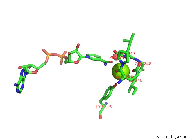
Magnesium Binding Sites:
The binding sites of Magnesium atom in the Pyld Holoenzyme
(pdb code 4j43). This binding sites where shown within
5.0 Angstroms radius around Magnesium atom.
In total 2 binding sites of Magnesium where determined in the Pyld Holoenzyme, PDB code: 4j43:
Jump to Magnesium binding site number: 1; 2;
In total 2 binding sites of Magnesium where determined in the Pyld Holoenzyme, PDB code: 4j43:
Jump to Magnesium binding site number: 1; 2;
Magnesium binding site 1 out of 2 in 4j43
Go back to
Magnesium binding site 1 out
of 2 in the Pyld Holoenzyme
Mono view
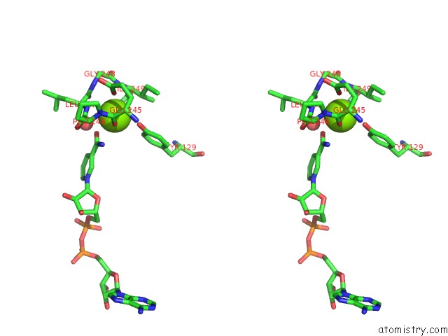
Stereo pair view
Mono view
Stereo pair view
A full contact list of Magnesium with other atoms in the Mg binding
site number 1 of Pyld Holoenzyme within 5.0Å range:
|
Magnesium binding site 2 out of 2 in 4j43
Go back to
Magnesium binding site 2 out
of 2 in the Pyld Holoenzyme

Mono view
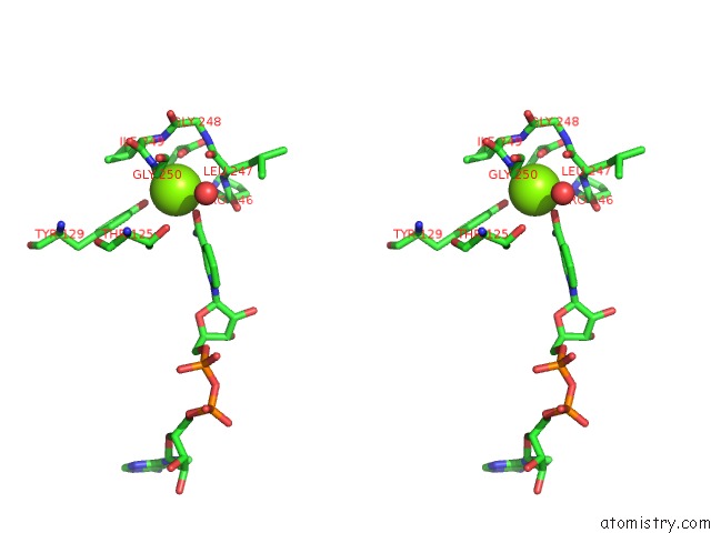
Stereo pair view
Mono view
Stereo pair view
A full contact list of Magnesium with other atoms in the Mg binding
site number 2 of Pyld Holoenzyme within 5.0Å range:
|
Reference:
F.Quitterer,
P.Beck,
A.Bacher,
M.Groll.
Structure and Reaction Mechanism of Pyrrolysine Synthase (Pyld). Angew.Chem.Int.Ed.Engl. V. 52 7033 2013.
ISSN: ISSN 1433-7851
PubMed: 23720358
DOI: 10.1002/ANIE.201301164
Page generated: Fri Aug 16 17:09:15 2024
ISSN: ISSN 1433-7851
PubMed: 23720358
DOI: 10.1002/ANIE.201301164
Last articles
Zn in 9J0NZn in 9J0O
Zn in 9J0P
Zn in 9FJX
Zn in 9EKB
Zn in 9C0F
Zn in 9CAH
Zn in 9CH0
Zn in 9CH3
Zn in 9CH1