Magnesium »
PDB 4j9l-4jjs »
4jdi »
Magnesium in PDB 4jdi: Crystal Structure of Serine/Threonine-Protein Kinase Pak 4 in Complex with Paktide S Peptide Substrate
Enzymatic activity of Crystal Structure of Serine/Threonine-Protein Kinase Pak 4 in Complex with Paktide S Peptide Substrate
All present enzymatic activity of Crystal Structure of Serine/Threonine-Protein Kinase Pak 4 in Complex with Paktide S Peptide Substrate:
2.7.11.1;
2.7.11.1;
Protein crystallography data
The structure of Crystal Structure of Serine/Threonine-Protein Kinase Pak 4 in Complex with Paktide S Peptide Substrate, PDB code: 4jdi
was solved by
B.H.Ha,
T.J.Boggon,
with X-Ray Crystallography technique. A brief refinement statistics is given in the table below:
| Resolution Low / High (Å) | 29.32 / 1.85 |
| Space group | P 41 21 2 |
| Cell size a, b, c (Å), α, β, γ (°) | 61.912, 61.912, 181.560, 90.00, 90.00, 90.00 |
| R / Rfree (%) | 19.2 / 23.3 |
Magnesium Binding Sites:
The binding sites of Magnesium atom in the Crystal Structure of Serine/Threonine-Protein Kinase Pak 4 in Complex with Paktide S Peptide Substrate
(pdb code 4jdi). This binding sites where shown within
5.0 Angstroms radius around Magnesium atom.
In total 2 binding sites of Magnesium where determined in the Crystal Structure of Serine/Threonine-Protein Kinase Pak 4 in Complex with Paktide S Peptide Substrate, PDB code: 4jdi:
Jump to Magnesium binding site number: 1; 2;
In total 2 binding sites of Magnesium where determined in the Crystal Structure of Serine/Threonine-Protein Kinase Pak 4 in Complex with Paktide S Peptide Substrate, PDB code: 4jdi:
Jump to Magnesium binding site number: 1; 2;
Magnesium binding site 1 out of 2 in 4jdi
Go back to
Magnesium binding site 1 out
of 2 in the Crystal Structure of Serine/Threonine-Protein Kinase Pak 4 in Complex with Paktide S Peptide Substrate
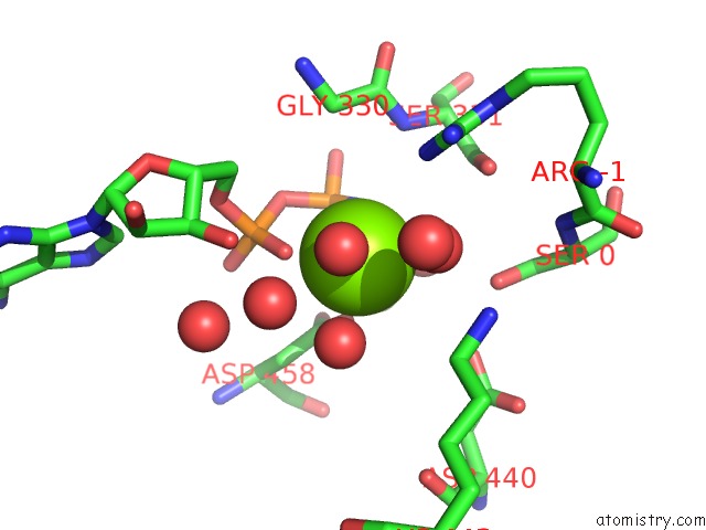
Mono view
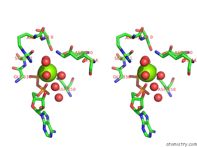
Stereo pair view
Mono view
Stereo pair view
A full contact list of Magnesium with other atoms in the Mg binding
site number 1 of Crystal Structure of Serine/Threonine-Protein Kinase Pak 4 in Complex with Paktide S Peptide Substrate within 5.0Å range:
|
Magnesium binding site 2 out of 2 in 4jdi
Go back to
Magnesium binding site 2 out
of 2 in the Crystal Structure of Serine/Threonine-Protein Kinase Pak 4 in Complex with Paktide S Peptide Substrate
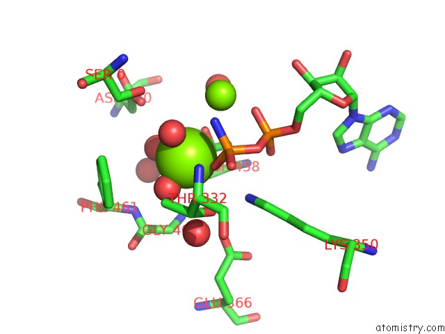
Mono view
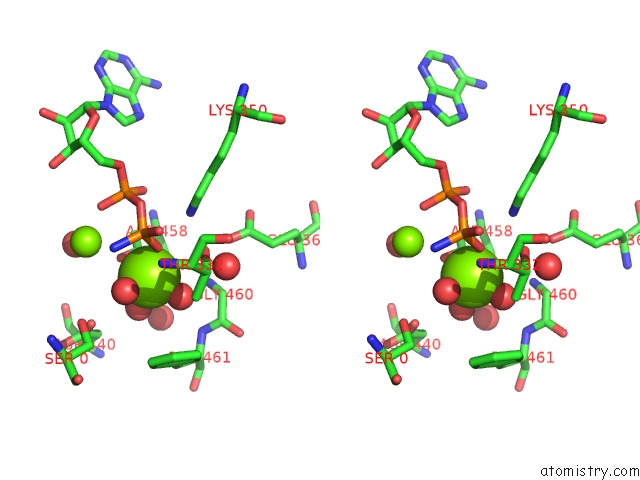
Stereo pair view
Mono view
Stereo pair view
A full contact list of Magnesium with other atoms in the Mg binding
site number 2 of Crystal Structure of Serine/Threonine-Protein Kinase Pak 4 in Complex with Paktide S Peptide Substrate within 5.0Å range:
|
Reference:
C.Chen,
B.H.Ha,
A.F.Thevenin,
H.J.Lou,
R.Zhang,
K.Y.Yip,
J.R.Peterson,
M.Gerstein,
P.M.Kim,
P.Filippakopoulos,
S.Knapp,
T.J.Boggon,
B.E.Turk.
Identification of A Major Determinant For Serine-Threonine Kinase Phosphoacceptor Specificity. Mol.Cell V. 53 140 2014.
ISSN: ISSN 1097-2765
PubMed: 24374310
DOI: 10.1016/J.MOLCEL.2013.11.013
Page generated: Mon Aug 11 14:41:20 2025
ISSN: ISSN 1097-2765
PubMed: 24374310
DOI: 10.1016/J.MOLCEL.2013.11.013
Last articles
Mg in 4LN7Mg in 4LMN
Mg in 4LLY
Mg in 4LLG
Mg in 4LK1
Mg in 4LK0
Mg in 4LJZ
Mg in 4LJY
Mg in 4LJA
Mg in 4LFC