Magnesium »
PDB 4jju-4jty »
4jqp »
Magnesium in PDB 4jqp: X-Ray Crystal Structure of A 4-Hydroxythreonine-4-Phosphate Dehydrogenase From Burkholderia Phymatum
Enzymatic activity of X-Ray Crystal Structure of A 4-Hydroxythreonine-4-Phosphate Dehydrogenase From Burkholderia Phymatum
All present enzymatic activity of X-Ray Crystal Structure of A 4-Hydroxythreonine-4-Phosphate Dehydrogenase From Burkholderia Phymatum:
1.1.1.262;
1.1.1.262;
Protein crystallography data
The structure of X-Ray Crystal Structure of A 4-Hydroxythreonine-4-Phosphate Dehydrogenase From Burkholderia Phymatum, PDB code: 4jqp
was solved by
Seattle Structural Genomics Center For Infectious Disease (Ssgcid),
with X-Ray Crystallography technique. A brief refinement statistics is given in the table below:
| Resolution Low / High (Å) | 48.91 / 1.65 |
| Space group | P 1 21 1 |
| Cell size a, b, c (Å), α, β, γ (°) | 48.980, 99.280, 71.030, 90.00, 93.92, 90.00 |
| R / Rfree (%) | 14.8 / 17.4 |
Other elements in 4jqp:
The structure of X-Ray Crystal Structure of A 4-Hydroxythreonine-4-Phosphate Dehydrogenase From Burkholderia Phymatum also contains other interesting chemical elements:
| Calcium | (Ca) | 2 atoms |
| Zinc | (Zn) | 2 atoms |
Magnesium Binding Sites:
The binding sites of Magnesium atom in the X-Ray Crystal Structure of A 4-Hydroxythreonine-4-Phosphate Dehydrogenase From Burkholderia Phymatum
(pdb code 4jqp). This binding sites where shown within
5.0 Angstroms radius around Magnesium atom.
In total 2 binding sites of Magnesium where determined in the X-Ray Crystal Structure of A 4-Hydroxythreonine-4-Phosphate Dehydrogenase From Burkholderia Phymatum, PDB code: 4jqp:
Jump to Magnesium binding site number: 1; 2;
In total 2 binding sites of Magnesium where determined in the X-Ray Crystal Structure of A 4-Hydroxythreonine-4-Phosphate Dehydrogenase From Burkholderia Phymatum, PDB code: 4jqp:
Jump to Magnesium binding site number: 1; 2;
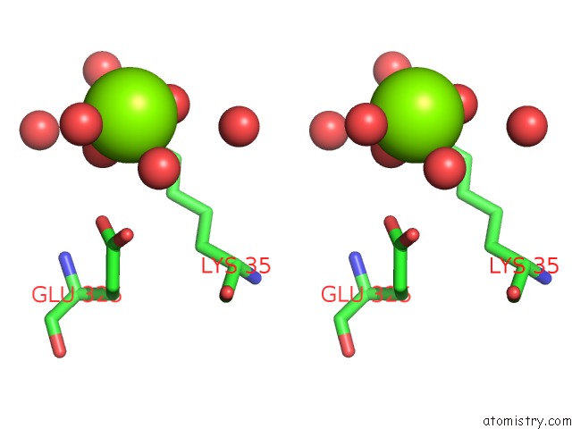
Magnesium binding site 1 out of 2 in 4jqp
Go back to
Magnesium binding site 1 out
of 2 in the X-Ray Crystal Structure of A 4-Hydroxythreonine-4-Phosphate Dehydrogenase From Burkholderia Phymatum

Mono view

Stereo pair view
Mono view
Stereo pair view
A full contact list of Magnesium with other atoms in the Mg binding
site number 1 of X-Ray Crystal Structure of A 4-Hydroxythreonine-4-Phosphate Dehydrogenase From Burkholderia Phymatum within 5.0Å range:
|
Magnesium binding site 2 out of 2 in 4jqp
Go back to
Magnesium binding site 2 out
of 2 in the X-Ray Crystal Structure of A 4-Hydroxythreonine-4-Phosphate Dehydrogenase From Burkholderia Phymatum

Mono view
Stereo pair view
Mono view
Stereo pair view
A full contact list of Magnesium with other atoms in the Mg binding
site number 2 of X-Ray Crystal Structure of A 4-Hydroxythreonine-4-Phosphate Dehydrogenase From Burkholderia Phymatum within 5.0Å range:
|
Reference:
J.W.Fairman,
M.C.Clifton,
T.E.Edwards,
D.Lorimer.
X-Ray Crystal Structure of A 4-Hydroxythreonine-4-Phosphate Dehydrogenase From Burkholderia Phymatum To Be Published.
Page generated: Mon Aug 11 17:16:36 2025
Last articles
Mg in 4S1LMg in 4S1K
Mg in 4S1I
Mg in 4S17
Mg in 4S1H
Mg in 4RYU
Mg in 4S0M
Mg in 4S04
Mg in 4S05
Mg in 4RXS