Magnesium »
PDB 4jty-4k5p »
4ju2 »
Magnesium in PDB 4ju2: Crystal Structure of Hcv NS5B Polymerase in Complex with Compound 12
Enzymatic activity of Crystal Structure of Hcv NS5B Polymerase in Complex with Compound 12
All present enzymatic activity of Crystal Structure of Hcv NS5B Polymerase in Complex with Compound 12:
2.7.7.48; 3.4.21.98; 3.6.1.15; 3.6.4.13;
2.7.7.48; 3.4.21.98; 3.6.1.15; 3.6.4.13;
Protein crystallography data
The structure of Crystal Structure of Hcv NS5B Polymerase in Complex with Compound 12, PDB code: 4ju2
was solved by
R.Coulombe,
with X-Ray Crystallography technique. A brief refinement statistics is given in the table below:
| Resolution Low / High (Å) | 50.00 / 2.70 |
| Space group | P 21 21 21 |
| Cell size a, b, c (Å), α, β, γ (°) | 105.540, 107.740, 135.820, 90.00, 90.00, 90.00 |
| R / Rfree (%) | n/a / n/a |
Other elements in 4ju2:
The structure of Crystal Structure of Hcv NS5B Polymerase in Complex with Compound 12 also contains other interesting chemical elements:
| Fluorine | (F) | 12 atoms |
Magnesium Binding Sites:
The binding sites of Magnesium atom in the Crystal Structure of Hcv NS5B Polymerase in Complex with Compound 12
(pdb code 4ju2). This binding sites where shown within
5.0 Angstroms radius around Magnesium atom.
In total 4 binding sites of Magnesium where determined in the Crystal Structure of Hcv NS5B Polymerase in Complex with Compound 12, PDB code: 4ju2:
Jump to Magnesium binding site number: 1; 2; 3; 4;
In total 4 binding sites of Magnesium where determined in the Crystal Structure of Hcv NS5B Polymerase in Complex with Compound 12, PDB code: 4ju2:
Jump to Magnesium binding site number: 1; 2; 3; 4;
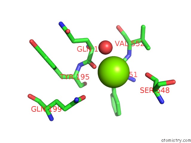
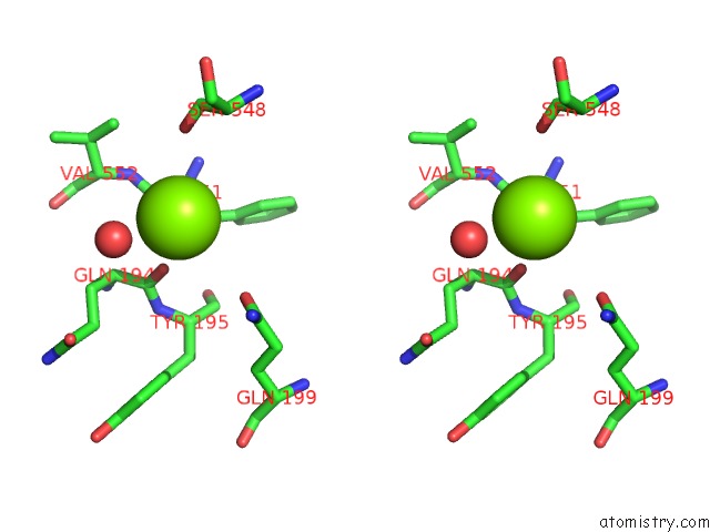
Magnesium binding site 1 out of 4 in 4ju2
Go back to
Magnesium binding site 1 out
of 4 in the Crystal Structure of Hcv NS5B Polymerase in Complex with Compound 12
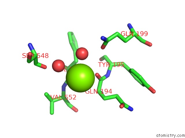
Mono view
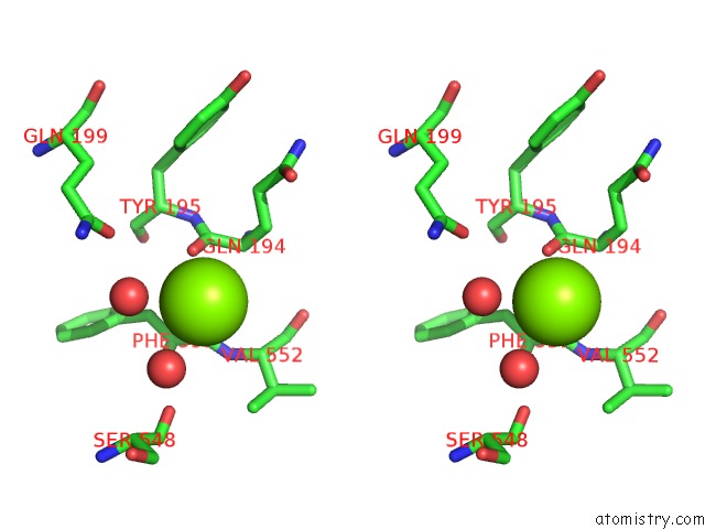
Stereo pair view
Mono view
Stereo pair view
A full contact list of Magnesium with other atoms in the Mg binding
site number 1 of Crystal Structure of Hcv NS5B Polymerase in Complex with Compound 12 within 5.0Å range:
|
Magnesium binding site 2 out of 4 in 4ju2
Go back to
Magnesium binding site 2 out
of 4 in the Crystal Structure of Hcv NS5B Polymerase in Complex with Compound 12
Mono view
Stereo pair view
Mono view
Stereo pair view
A full contact list of Magnesium with other atoms in the Mg binding
site number 2 of Crystal Structure of Hcv NS5B Polymerase in Complex with Compound 12 within 5.0Å range:
|
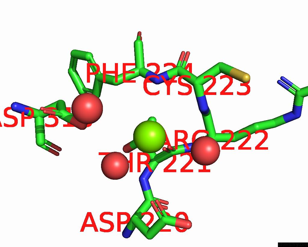
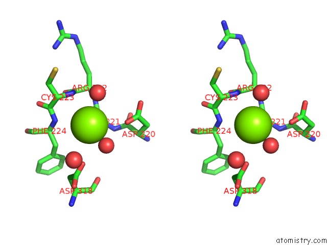
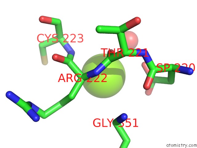
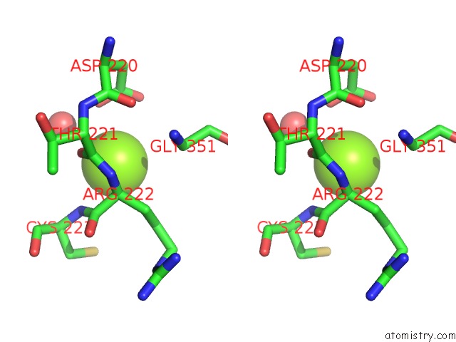
Magnesium binding site 3 out of 4 in 4ju2
Go back to
Magnesium binding site 3 out
of 4 in the Crystal Structure of Hcv NS5B Polymerase in Complex with Compound 12
Mono view
Stereo pair view
Mono view
Stereo pair view
A full contact list of Magnesium with other atoms in the Mg binding
site number 3 of Crystal Structure of Hcv NS5B Polymerase in Complex with Compound 12 within 5.0Å range:
|
Magnesium binding site 4 out of 4 in 4ju2
Go back to
Magnesium binding site 4 out
of 4 in the Crystal Structure of Hcv NS5B Polymerase in Complex with Compound 12
Mono view
Stereo pair view
Mono view
Stereo pair view
A full contact list of Magnesium with other atoms in the Mg binding
site number 4 of Crystal Structure of Hcv NS5B Polymerase in Complex with Compound 12 within 5.0Å range:
|
Reference:
O.Hucke,
R.Coulombe,
P.Bonneau,
M.Bertrand-Laperle,
C.Brochu,
J.Gillard,
M.A.Joly,
S.Landry,
O.Lepage,
M.Llinas-Brunet,
M.Pesant,
M.Poirier,
M.Poirier,
G.Mckercher,
M.Marquis,
G.Kukolj,
P.L.Beaulieu,
T.A.Stammers.
Molecular Dynamics Simulations and Structure-Based Rational Design Lead to Allosteric Hcv NS5B Polymerase Thumb Pocket 2 Inhibitor with Picomolar Cellular Replicon Potency. J.Med.Chem. V. 57 1932 2014.
ISSN: ISSN 0022-2623
PubMed: 23773186
DOI: 10.1021/JM4004522
Page generated: Sat Aug 17 03:17:06 2024
ISSN: ISSN 0022-2623
PubMed: 23773186
DOI: 10.1021/JM4004522
Last articles
Zn in 9J0NZn in 9J0O
Zn in 9J0P
Zn in 9FJX
Zn in 9EKB
Zn in 9C0F
Zn in 9CAH
Zn in 9CH0
Zn in 9CH3
Zn in 9CH1