Magnesium »
PDB 4jtz-4k6e »
4jx5 »
Magnesium in PDB 4jx5: Structure of the Carboxyl Transferase Domain From Rhizobium Etli Pyruvate Carboxylase with Pyruvate
Enzymatic activity of Structure of the Carboxyl Transferase Domain From Rhizobium Etli Pyruvate Carboxylase with Pyruvate
All present enzymatic activity of Structure of the Carboxyl Transferase Domain From Rhizobium Etli Pyruvate Carboxylase with Pyruvate:
6.4.1.1;
6.4.1.1;
Protein crystallography data
The structure of Structure of the Carboxyl Transferase Domain From Rhizobium Etli Pyruvate Carboxylase with Pyruvate, PDB code: 4jx5
was solved by
A.D.Lietzan,
M.St Maurice,
with X-Ray Crystallography technique. A brief refinement statistics is given in the table below:
| Resolution Low / High (Å) | 46.88 / 2.55 |
| Space group | P 21 21 21 |
| Cell size a, b, c (Å), α, β, γ (°) | 85.130, 157.253, 245.320, 90.00, 90.00, 90.00 |
| R / Rfree (%) | 18.7 / 23.2 |
Other elements in 4jx5:
The structure of Structure of the Carboxyl Transferase Domain From Rhizobium Etli Pyruvate Carboxylase with Pyruvate also contains other interesting chemical elements:
| Chlorine | (Cl) | 4 atoms |
| Zinc | (Zn) | 4 atoms |
Magnesium Binding Sites:
The binding sites of Magnesium atom in the Structure of the Carboxyl Transferase Domain From Rhizobium Etli Pyruvate Carboxylase with Pyruvate
(pdb code 4jx5). This binding sites where shown within
5.0 Angstroms radius around Magnesium atom.
In total 4 binding sites of Magnesium where determined in the Structure of the Carboxyl Transferase Domain From Rhizobium Etli Pyruvate Carboxylase with Pyruvate, PDB code: 4jx5:
Jump to Magnesium binding site number: 1; 2; 3; 4;
In total 4 binding sites of Magnesium where determined in the Structure of the Carboxyl Transferase Domain From Rhizobium Etli Pyruvate Carboxylase with Pyruvate, PDB code: 4jx5:
Jump to Magnesium binding site number: 1; 2; 3; 4;
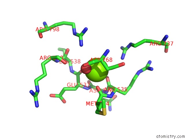
Magnesium binding site 1 out of 4 in 4jx5
Go back to
Magnesium binding site 1 out
of 4 in the Structure of the Carboxyl Transferase Domain From Rhizobium Etli Pyruvate Carboxylase with Pyruvate
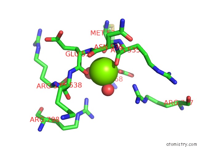
Mono view
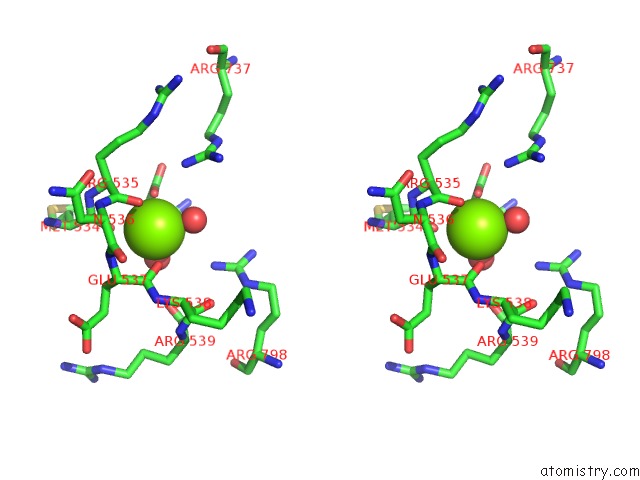
Stereo pair view
Mono view
Stereo pair view
A full contact list of Magnesium with other atoms in the Mg binding
site number 1 of Structure of the Carboxyl Transferase Domain From Rhizobium Etli Pyruvate Carboxylase with Pyruvate within 5.0Å range:
|
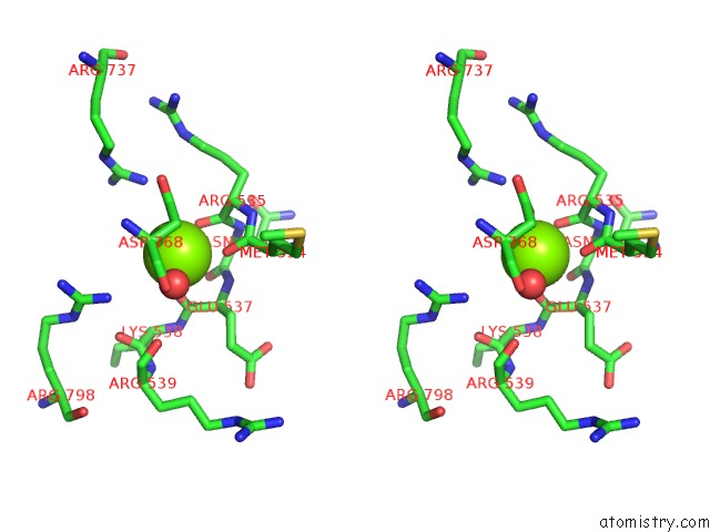
Magnesium binding site 2 out of 4 in 4jx5
Go back to
Magnesium binding site 2 out
of 4 in the Structure of the Carboxyl Transferase Domain From Rhizobium Etli Pyruvate Carboxylase with Pyruvate
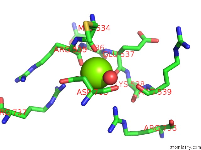
Mono view
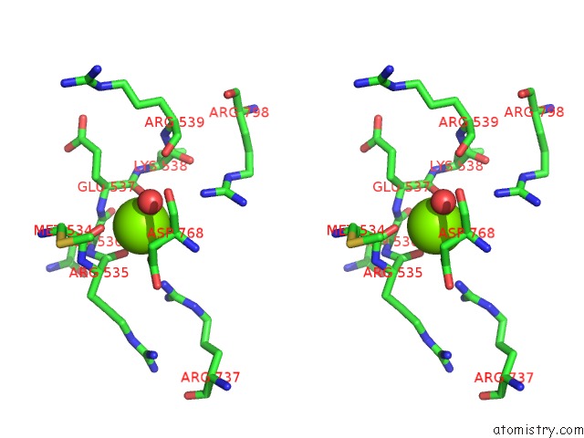
Stereo pair view
Mono view
Stereo pair view
A full contact list of Magnesium with other atoms in the Mg binding
site number 2 of Structure of the Carboxyl Transferase Domain From Rhizobium Etli Pyruvate Carboxylase with Pyruvate within 5.0Å range:
|
Magnesium binding site 3 out of 4 in 4jx5
Go back to
Magnesium binding site 3 out
of 4 in the Structure of the Carboxyl Transferase Domain From Rhizobium Etli Pyruvate Carboxylase with Pyruvate

Mono view

Stereo pair view
Mono view
Stereo pair view
A full contact list of Magnesium with other atoms in the Mg binding
site number 3 of Structure of the Carboxyl Transferase Domain From Rhizobium Etli Pyruvate Carboxylase with Pyruvate within 5.0Å range:
|
Magnesium binding site 4 out of 4 in 4jx5
Go back to
Magnesium binding site 4 out
of 4 in the Structure of the Carboxyl Transferase Domain From Rhizobium Etli Pyruvate Carboxylase with Pyruvate
Mono view
Stereo pair view
Mono view
Stereo pair view
A full contact list of Magnesium with other atoms in the Mg binding
site number 4 of Structure of the Carboxyl Transferase Domain From Rhizobium Etli Pyruvate Carboxylase with Pyruvate within 5.0Å range:
|
Reference:
A.D.Lietzan,
M.St Maurice.
A Substrate-Induced Biotin Binding Pocket in the Carboxyltransferase Domain of Pyruvate Carboxylase. J.Biol.Chem. V. 288 19915 2013.
ISSN: ISSN 0021-9258
PubMed: 23698000
DOI: 10.1074/JBC.M113.477828
Page generated: Mon Aug 11 17:21:35 2025
ISSN: ISSN 0021-9258
PubMed: 23698000
DOI: 10.1074/JBC.M113.477828
Last articles
Mg in 4W5OMg in 4W5J
Mg in 4W5N
Mg in 4V2I
Mg in 4V3R
Mg in 4V26
Mg in 4V2G
Mg in 4V1T
Mg in 4V25
Mg in 4V1V