Magnesium »
PDB 4k6e-4kft »
4k6o »
Magnesium in PDB 4k6o: X-Ray Structure Uridine Phosphorylase From Vibrio Cholerae in Complex with 6-Methyluracil at 1.17 A Resolution
Enzymatic activity of X-Ray Structure Uridine Phosphorylase From Vibrio Cholerae in Complex with 6-Methyluracil at 1.17 A Resolution
All present enzymatic activity of X-Ray Structure Uridine Phosphorylase From Vibrio Cholerae in Complex with 6-Methyluracil at 1.17 A Resolution:
2.4.2.3;
2.4.2.3;
Protein crystallography data
The structure of X-Ray Structure Uridine Phosphorylase From Vibrio Cholerae in Complex with 6-Methyluracil at 1.17 A Resolution, PDB code: 4k6o
was solved by
I.I.Prokofev,
A.A.Lashkov,
A.G.Gabdoulkhakov,
C.Betzel,
A.M.Mikhailov,
with X-Ray Crystallography technique. A brief refinement statistics is given in the table below:
| Resolution Low / High (Å) | 40.28 / 1.17 |
| Space group | P 1 21 1 |
| Cell size a, b, c (Å), α, β, γ (°) | 93.012, 97.100, 93.017, 90.00, 119.99, 90.00 |
| R / Rfree (%) | 10.8 / 12.8 |
Other elements in 4k6o:
The structure of X-Ray Structure Uridine Phosphorylase From Vibrio Cholerae in Complex with 6-Methyluracil at 1.17 A Resolution also contains other interesting chemical elements:
| Chlorine | (Cl) | 12 atoms |
| Sodium | (Na) | 4 atoms |
Magnesium Binding Sites:
Pages:
>>> Page 1 <<< Page 2, Binding sites: 11 - 16;Binding sites:
The binding sites of Magnesium atom in the X-Ray Structure Uridine Phosphorylase From Vibrio Cholerae in Complex with 6-Methyluracil at 1.17 A Resolution (pdb code 4k6o). This binding sites where shown within 5.0 Angstroms radius around Magnesium atom.In total 16 binding sites of Magnesium where determined in the X-Ray Structure Uridine Phosphorylase From Vibrio Cholerae in Complex with 6-Methyluracil at 1.17 A Resolution, PDB code: 4k6o:
Jump to Magnesium binding site number: 1; 2; 3; 4; 5; 6; 7; 8; 9; 10;
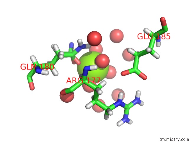
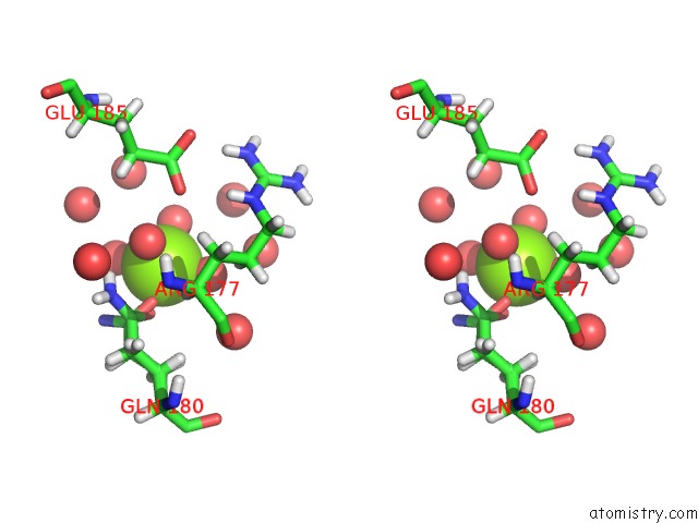
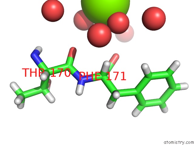
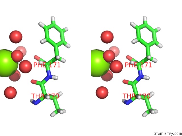
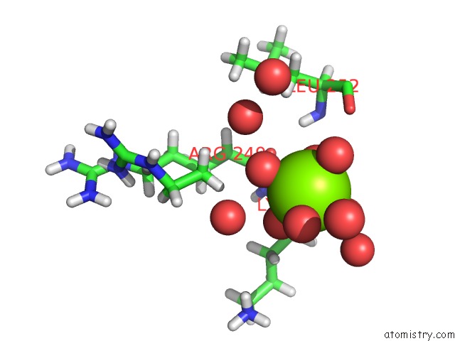
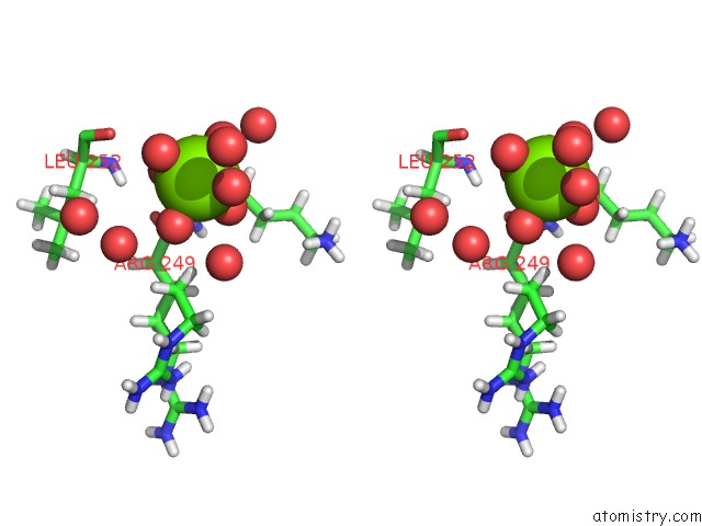
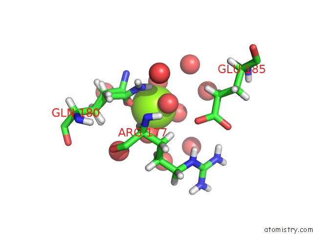
Magnesium binding site 1 out of 16 in 4k6o
Go back to
Magnesium binding site 1 out
of 16 in the X-Ray Structure Uridine Phosphorylase From Vibrio Cholerae in Complex with 6-Methyluracil at 1.17 A Resolution
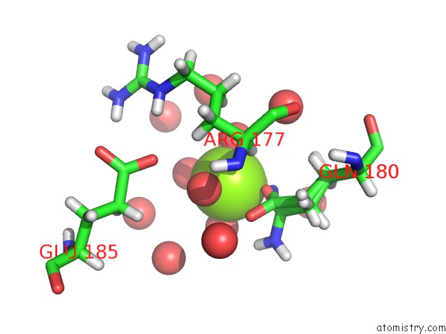
Mono view
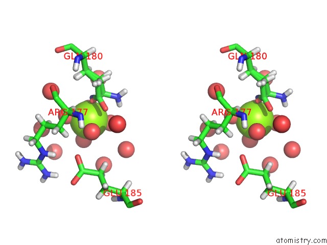
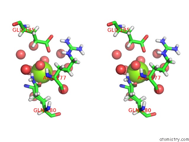
Stereo pair view
Mono view
Stereo pair view
A full contact list of Magnesium with other atoms in the Mg binding
site number 1 of X-Ray Structure Uridine Phosphorylase From Vibrio Cholerae in Complex with 6-Methyluracil at 1.17 A Resolution within 5.0Å range:
|
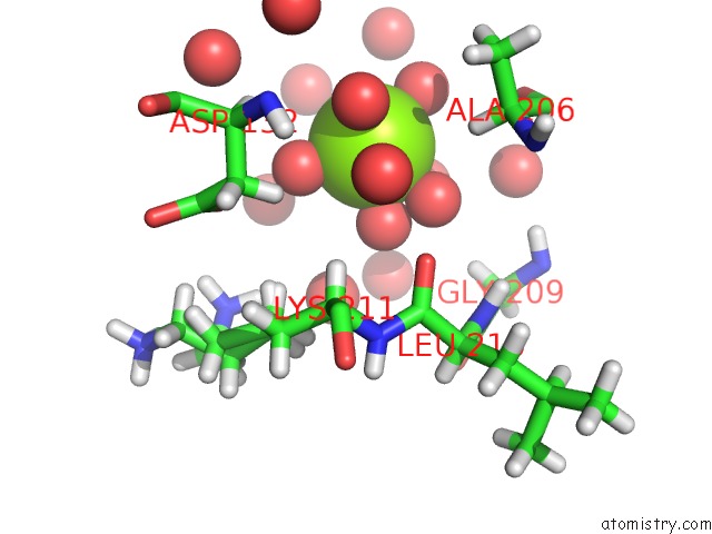
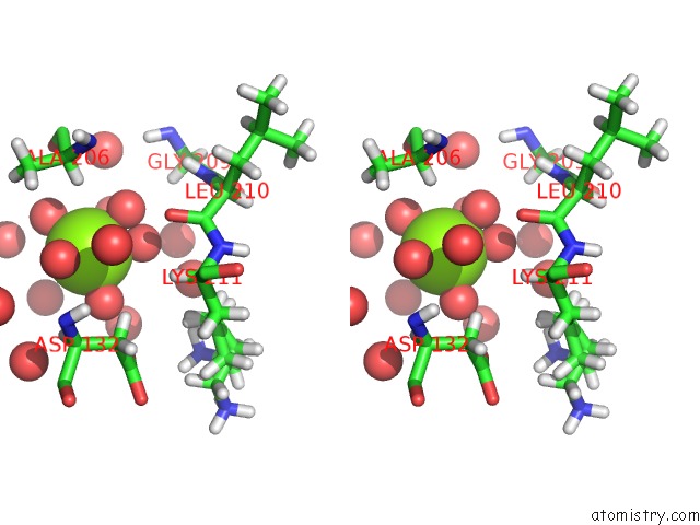
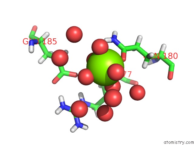
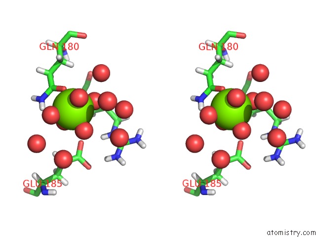
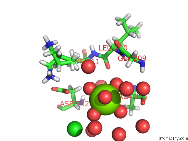
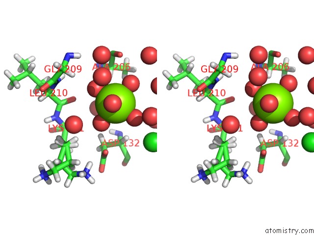
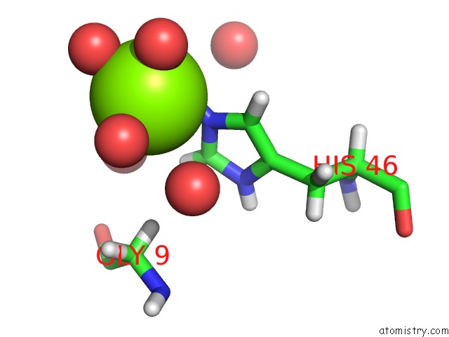
Magnesium binding site 2 out of 16 in 4k6o
Go back to
Magnesium binding site 2 out
of 16 in the X-Ray Structure Uridine Phosphorylase From Vibrio Cholerae in Complex with 6-Methyluracil at 1.17 A Resolution
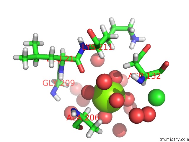
Mono view
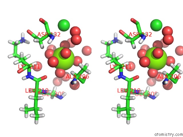
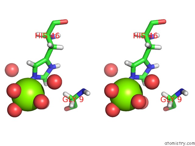
Stereo pair view
Mono view
Stereo pair view
A full contact list of Magnesium with other atoms in the Mg binding
site number 2 of X-Ray Structure Uridine Phosphorylase From Vibrio Cholerae in Complex with 6-Methyluracil at 1.17 A Resolution within 5.0Å range:
|
Magnesium binding site 3 out of 16 in 4k6o
Go back to
Magnesium binding site 3 out
of 16 in the X-Ray Structure Uridine Phosphorylase From Vibrio Cholerae in Complex with 6-Methyluracil at 1.17 A Resolution
Mono view
Stereo pair view
Mono view
Stereo pair view
A full contact list of Magnesium with other atoms in the Mg binding
site number 3 of X-Ray Structure Uridine Phosphorylase From Vibrio Cholerae in Complex with 6-Methyluracil at 1.17 A Resolution within 5.0Å range:
|
Magnesium binding site 4 out of 16 in 4k6o
Go back to
Magnesium binding site 4 out
of 16 in the X-Ray Structure Uridine Phosphorylase From Vibrio Cholerae in Complex with 6-Methyluracil at 1.17 A Resolution
Mono view
Stereo pair view
Mono view
Stereo pair view
A full contact list of Magnesium with other atoms in the Mg binding
site number 4 of X-Ray Structure Uridine Phosphorylase From Vibrio Cholerae in Complex with 6-Methyluracil at 1.17 A Resolution within 5.0Å range:
|
Magnesium binding site 5 out of 16 in 4k6o
Go back to
Magnesium binding site 5 out
of 16 in the X-Ray Structure Uridine Phosphorylase From Vibrio Cholerae in Complex with 6-Methyluracil at 1.17 A Resolution
Mono view
Stereo pair view
Mono view
Stereo pair view
A full contact list of Magnesium with other atoms in the Mg binding
site number 5 of X-Ray Structure Uridine Phosphorylase From Vibrio Cholerae in Complex with 6-Methyluracil at 1.17 A Resolution within 5.0Å range:
|
Magnesium binding site 6 out of 16 in 4k6o
Go back to
Magnesium binding site 6 out
of 16 in the X-Ray Structure Uridine Phosphorylase From Vibrio Cholerae in Complex with 6-Methyluracil at 1.17 A Resolution
Mono view
Stereo pair view
Mono view
Stereo pair view
A full contact list of Magnesium with other atoms in the Mg binding
site number 6 of X-Ray Structure Uridine Phosphorylase From Vibrio Cholerae in Complex with 6-Methyluracil at 1.17 A Resolution within 5.0Å range:
|
Magnesium binding site 7 out of 16 in 4k6o
Go back to
Magnesium binding site 7 out
of 16 in the X-Ray Structure Uridine Phosphorylase From Vibrio Cholerae in Complex with 6-Methyluracil at 1.17 A Resolution
Mono view
Stereo pair view
Mono view
Stereo pair view
A full contact list of Magnesium with other atoms in the Mg binding
site number 7 of X-Ray Structure Uridine Phosphorylase From Vibrio Cholerae in Complex with 6-Methyluracil at 1.17 A Resolution within 5.0Å range:
|
Magnesium binding site 8 out of 16 in 4k6o
Go back to
Magnesium binding site 8 out
of 16 in the X-Ray Structure Uridine Phosphorylase From Vibrio Cholerae in Complex with 6-Methyluracil at 1.17 A Resolution
Mono view
Stereo pair view
Mono view
Stereo pair view
A full contact list of Magnesium with other atoms in the Mg binding
site number 8 of X-Ray Structure Uridine Phosphorylase From Vibrio Cholerae in Complex with 6-Methyluracil at 1.17 A Resolution within 5.0Å range:
|
Magnesium binding site 9 out of 16 in 4k6o
Go back to
Magnesium binding site 9 out
of 16 in the X-Ray Structure Uridine Phosphorylase From Vibrio Cholerae in Complex with 6-Methyluracil at 1.17 A Resolution
Mono view
Stereo pair view
Mono view
Stereo pair view
A full contact list of Magnesium with other atoms in the Mg binding
site number 9 of X-Ray Structure Uridine Phosphorylase From Vibrio Cholerae in Complex with 6-Methyluracil at 1.17 A Resolution within 5.0Å range:
|
Magnesium binding site 10 out of 16 in 4k6o
Go back to
Magnesium binding site 10 out
of 16 in the X-Ray Structure Uridine Phosphorylase From Vibrio Cholerae in Complex with 6-Methyluracil at 1.17 A Resolution
Mono view
Stereo pair view
Mono view
Stereo pair view
A full contact list of Magnesium with other atoms in the Mg binding
site number 10 of X-Ray Structure Uridine Phosphorylase From Vibrio Cholerae in Complex with 6-Methyluracil at 1.17 A Resolution within 5.0Å range:
|
Reference:
I.I.Prokofev,
A.A.Lashkov,
A.G.Gabdoulkhakov,
M.V.Dontsova,
T.A.Seregina,
A.S.Mironov,
C.Betzel,
A.M.Mikhailov.
Crystallization and Preliminary X-Ray Study of Vibrio Cholerae Uridine Phosphorylase in Complex with 6-Methyluracil Acta Crystallogr.,Sect.F V. 70 60 2014.
ISSN: ESSN 1744-3091
DOI: 10.1107/S2053230X13031877
Page generated: Sat Aug 17 03:27:27 2024
ISSN: ESSN 1744-3091
DOI: 10.1107/S2053230X13031877
Last articles
Zn in 9JYWZn in 9IR4
Zn in 9IR3
Zn in 9GMX
Zn in 9GMW
Zn in 9JEJ
Zn in 9ERF
Zn in 9ERE
Zn in 9EGV
Zn in 9EGW