Magnesium »
PDB 4k6o-4kfu »
4kb0 »
Magnesium in PDB 4kb0: Crystal Structure of Rnase T in Complex with A Bluge Dna (Two Nucleotide Insertion Cc )
Protein crystallography data
The structure of Crystal Structure of Rnase T in Complex with A Bluge Dna (Two Nucleotide Insertion Cc ), PDB code: 4kb0
was solved by
Y.-Y.Hsiao,
H.S.Yuan,
with X-Ray Crystallography technique. A brief refinement statistics is given in the table below:
| Resolution Low / High (Å) | 29.80 / 2.00 |
| Space group | P 1 21 1 |
| Cell size a, b, c (Å), α, β, γ (°) | 60.257, 81.944, 73.444, 90.00, 105.46, 90.00 |
| R / Rfree (%) | 18.9 / 21.1 |
Magnesium Binding Sites:
The binding sites of Magnesium atom in the Crystal Structure of Rnase T in Complex with A Bluge Dna (Two Nucleotide Insertion Cc )
(pdb code 4kb0). This binding sites where shown within
5.0 Angstroms radius around Magnesium atom.
In total 4 binding sites of Magnesium where determined in the Crystal Structure of Rnase T in Complex with A Bluge Dna (Two Nucleotide Insertion Cc ), PDB code: 4kb0:
Jump to Magnesium binding site number: 1; 2; 3; 4;
In total 4 binding sites of Magnesium where determined in the Crystal Structure of Rnase T in Complex with A Bluge Dna (Two Nucleotide Insertion Cc ), PDB code: 4kb0:
Jump to Magnesium binding site number: 1; 2; 3; 4;
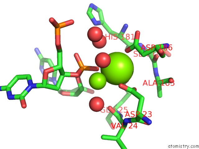
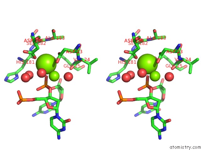
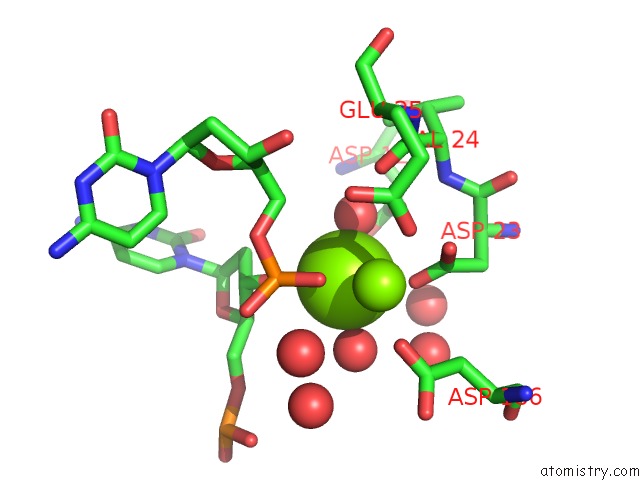
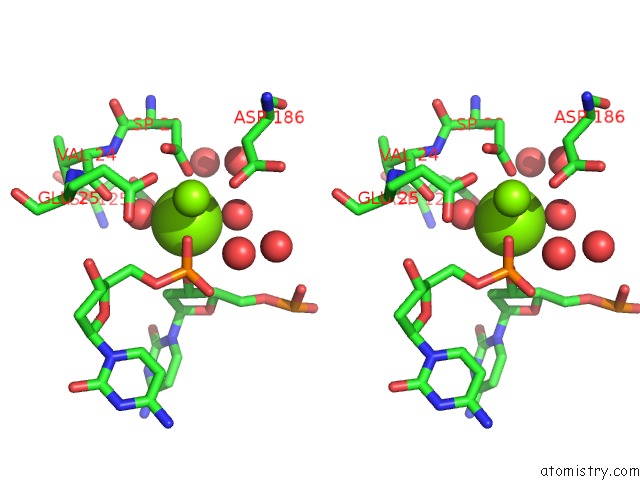
Magnesium binding site 1 out of 4 in 4kb0
Go back to
Magnesium binding site 1 out
of 4 in the Crystal Structure of Rnase T in Complex with A Bluge Dna (Two Nucleotide Insertion Cc )
Mono view
Stereo pair view
Mono view
Stereo pair view
A full contact list of Magnesium with other atoms in the Mg binding
site number 1 of Crystal Structure of Rnase T in Complex with A Bluge Dna (Two Nucleotide Insertion Cc ) within 5.0Å range:
|
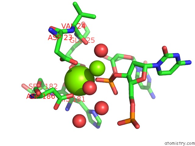
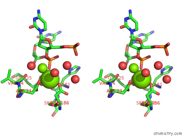
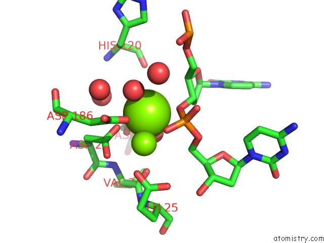
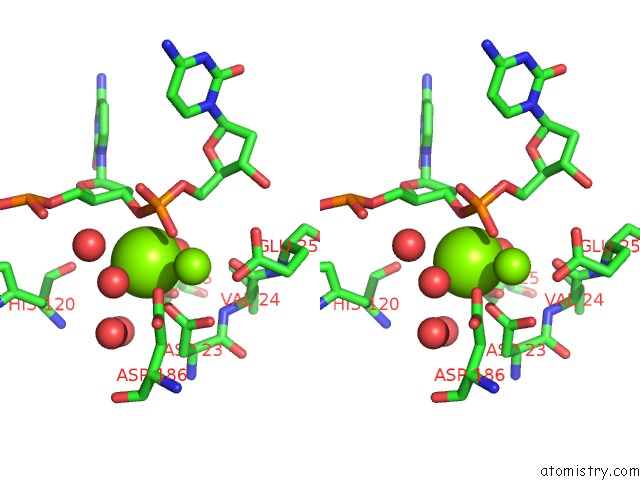
Magnesium binding site 2 out of 4 in 4kb0
Go back to
Magnesium binding site 2 out
of 4 in the Crystal Structure of Rnase T in Complex with A Bluge Dna (Two Nucleotide Insertion Cc )
Mono view
Stereo pair view
Mono view
Stereo pair view
A full contact list of Magnesium with other atoms in the Mg binding
site number 2 of Crystal Structure of Rnase T in Complex with A Bluge Dna (Two Nucleotide Insertion Cc ) within 5.0Å range:
|
Magnesium binding site 3 out of 4 in 4kb0
Go back to
Magnesium binding site 3 out
of 4 in the Crystal Structure of Rnase T in Complex with A Bluge Dna (Two Nucleotide Insertion Cc )
Mono view
Stereo pair view
Mono view
Stereo pair view
A full contact list of Magnesium with other atoms in the Mg binding
site number 3 of Crystal Structure of Rnase T in Complex with A Bluge Dna (Two Nucleotide Insertion Cc ) within 5.0Å range:
|
Magnesium binding site 4 out of 4 in 4kb0
Go back to
Magnesium binding site 4 out
of 4 in the Crystal Structure of Rnase T in Complex with A Bluge Dna (Two Nucleotide Insertion Cc )
Mono view
Stereo pair view
Mono view
Stereo pair view
A full contact list of Magnesium with other atoms in the Mg binding
site number 4 of Crystal Structure of Rnase T in Complex with A Bluge Dna (Two Nucleotide Insertion Cc ) within 5.0Å range:
|
Reference:
Y.Y.Hsiao,
W.H.Fang,
C.C.Lee,
Y.P.Chen,
H.S.Yuan.
Structural Insights Into Dna Repair By Rnase T--An Exonuclease Processing 3' End of Structured Dna in Repair Pathways. Plos Biol. V. 12 01803 2014.
ISSN: ISSN 1544-9173
PubMed: 24594808
DOI: 10.1371/JOURNAL.PBIO.1001803
Page generated: Sat Aug 17 03:32:15 2024
ISSN: ISSN 1544-9173
PubMed: 24594808
DOI: 10.1371/JOURNAL.PBIO.1001803
Last articles
Zn in 9MJ5Zn in 9HNW
Zn in 9G0L
Zn in 9FNE
Zn in 9DZN
Zn in 9E0I
Zn in 9D32
Zn in 9DAK
Zn in 8ZXC
Zn in 8ZUF