Magnesium in PDB 4kb1: Crystal Structure of Rnase T in Complex with A Bluge Dna (Two Nucleotide Insertion Ct )
Protein crystallography data
The structure of Crystal Structure of Rnase T in Complex with A Bluge Dna (Two Nucleotide Insertion Ct ), PDB code: 4kb1
was solved by
Y.-Y.Hsiao,
H.S.Yuan,
with X-Ray Crystallography technique. A brief refinement statistics is given in the table below:
Resolution Low / High (Å)
|
28.62 /
1.80
|
Space group
|
P 1 21 1
|
Cell size a, b, c (Å), α, β, γ (°)
|
60.556,
81.844,
73.744,
90.00,
105.71,
90.00
|
R / Rfree (%)
|
18.3 /
19.9
|
Magnesium Binding Sites:
The binding sites of Magnesium atom in the Crystal Structure of Rnase T in Complex with A Bluge Dna (Two Nucleotide Insertion Ct )
(pdb code 4kb1). This binding sites where shown within
5.0 Angstroms radius around Magnesium atom.
In total 4 binding sites of Magnesium where determined in the
Crystal Structure of Rnase T in Complex with A Bluge Dna (Two Nucleotide Insertion Ct ), PDB code: 4kb1:
Jump to Magnesium binding site number:
1;
2;
3;
4;
Magnesium binding site 1 out
of 4 in 4kb1
Go back to
Magnesium Binding Sites List in 4kb1
Magnesium binding site 1 out
of 4 in the Crystal Structure of Rnase T in Complex with A Bluge Dna (Two Nucleotide Insertion Ct )
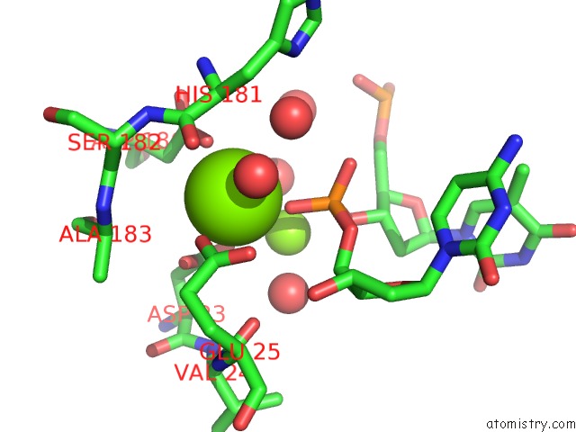
Mono view
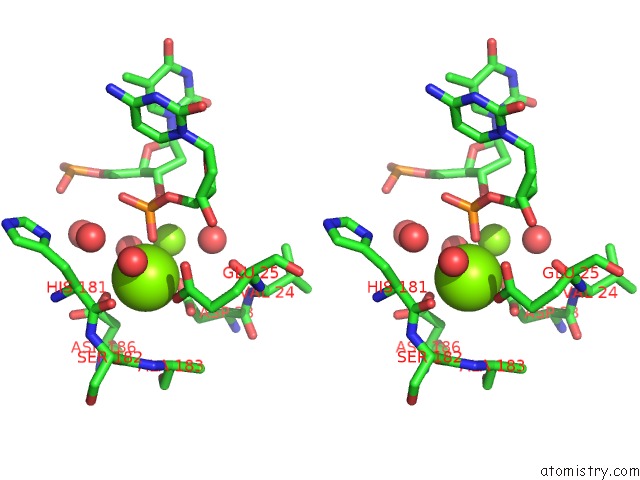
Stereo pair view
|
A full contact list of Magnesium with other atoms in the Mg binding
site number 1 of Crystal Structure of Rnase T in Complex with A Bluge Dna (Two Nucleotide Insertion Ct ) within 5.0Å range:
probe
|
atom
|
residue
|
distance (Å)
|
B
|
Occ
|
A:Mg301
b:26.3
occ:1.00
|
OP1
|
C:DC18
|
2.1
|
17.0
|
1.0
|
OD2
|
A:ASP186
|
2.3
|
20.9
|
1.0
|
OE1
|
A:GLU25
|
2.4
|
29.5
|
1.0
|
O
|
A:HOH502
|
2.5
|
36.3
|
1.0
|
OD1
|
A:ASP23
|
3.1
|
30.4
|
1.0
|
OD2
|
A:ASP23
|
3.2
|
27.3
|
1.0
|
CD
|
A:GLU25
|
3.3
|
25.5
|
1.0
|
CG
|
A:ASP186
|
3.4
|
25.2
|
1.0
|
O
|
C:HOH209
|
3.4
|
27.1
|
1.0
|
MG
|
C:MG101
|
3.5
|
19.4
|
1.0
|
CG
|
A:ASP23
|
3.5
|
21.0
|
1.0
|
P
|
C:DC18
|
3.5
|
16.2
|
1.0
|
OE2
|
A:GLU25
|
3.5
|
18.2
|
1.0
|
CB
|
A:ASP186
|
3.8
|
22.1
|
1.0
|
O
|
A:HOH424
|
3.9
|
20.6
|
1.0
|
CA
|
A:HIS181
|
4.2
|
33.1
|
1.0
|
C
|
A:HIS181
|
4.2
|
29.5
|
1.0
|
OP2
|
C:DC18
|
4.3
|
16.5
|
1.0
|
O5'
|
C:DC18
|
4.3
|
17.6
|
1.0
|
O
|
A:SER182
|
4.4
|
21.0
|
1.0
|
O
|
A:HIS181
|
4.4
|
30.1
|
1.0
|
O
|
A:HOH415
|
4.4
|
17.9
|
1.0
|
O3'
|
C:DT17
|
4.5
|
13.6
|
1.0
|
OD1
|
A:ASP186
|
4.5
|
20.1
|
1.0
|
O
|
A:VAL24
|
4.5
|
16.2
|
1.0
|
C
|
A:SER182
|
4.5
|
27.4
|
1.0
|
C5'
|
C:DC18
|
4.6
|
14.9
|
1.0
|
CA
|
A:ALA183
|
4.6
|
23.5
|
1.0
|
N
|
A:SER182
|
4.7
|
29.8
|
1.0
|
CG
|
A:GLU25
|
4.7
|
21.5
|
1.0
|
N
|
A:ALA183
|
4.7
|
23.3
|
1.0
|
O
|
C:HOH202
|
4.8
|
18.3
|
1.0
|
N
|
A:HIS181
|
4.9
|
34.2
|
1.0
|
CB
|
A:ASP23
|
5.0
|
16.5
|
1.0
|
|
Magnesium binding site 2 out
of 4 in 4kb1
Go back to
Magnesium Binding Sites List in 4kb1
Magnesium binding site 2 out
of 4 in the Crystal Structure of Rnase T in Complex with A Bluge Dna (Two Nucleotide Insertion Ct )
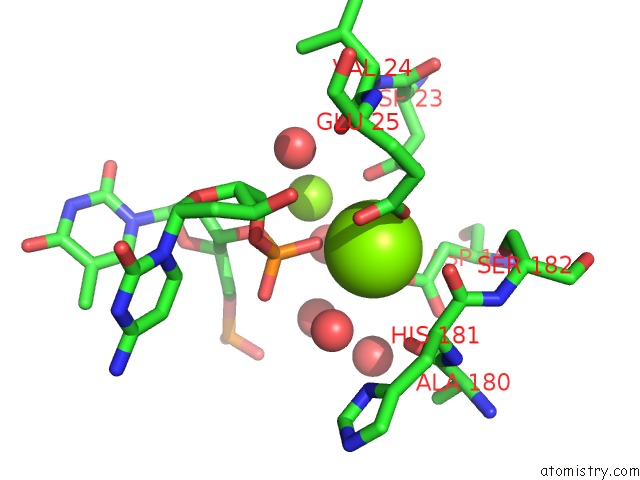
Mono view
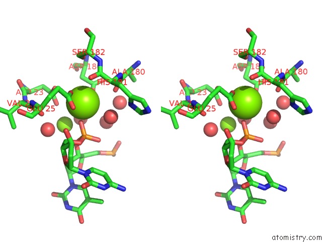
Stereo pair view
|
A full contact list of Magnesium with other atoms in the Mg binding
site number 2 of Crystal Structure of Rnase T in Complex with A Bluge Dna (Two Nucleotide Insertion Ct ) within 5.0Å range:
probe
|
atom
|
residue
|
distance (Å)
|
B
|
Occ
|
B:Mg301
b:30.8
occ:1.00
|
OP1
|
D:DC18
|
2.1
|
17.6
|
1.0
|
OE1
|
B:GLU25
|
2.2
|
31.4
|
1.0
|
OD2
|
B:ASP186
|
2.2
|
35.6
|
1.0
|
CD
|
B:GLU25
|
3.1
|
28.4
|
1.0
|
O
|
D:HOH212
|
3.3
|
39.1
|
1.0
|
OE2
|
B:GLU25
|
3.3
|
23.4
|
1.0
|
CG
|
B:ASP186
|
3.3
|
33.6
|
1.0
|
OD2
|
B:ASP23
|
3.4
|
24.7
|
1.0
|
P
|
D:DC18
|
3.5
|
16.4
|
1.0
|
O
|
B:ALA180
|
3.6
|
57.5
|
1.0
|
OD1
|
B:ASP23
|
3.7
|
18.6
|
1.0
|
MG
|
D:MG101
|
3.8
|
17.4
|
1.0
|
CG
|
B:ASP23
|
3.9
|
23.3
|
1.0
|
CB
|
B:ASP186
|
4.0
|
19.3
|
1.0
|
OP2
|
D:DC18
|
4.2
|
19.0
|
1.0
|
O
|
B:HOH424
|
4.2
|
19.3
|
1.0
|
O5'
|
D:DC18
|
4.2
|
14.5
|
1.0
|
CA
|
B:HIS181
|
4.2
|
44.0
|
1.0
|
O
|
B:HIS181
|
4.2
|
50.4
|
1.0
|
OD1
|
B:ASP186
|
4.3
|
33.6
|
1.0
|
C
|
B:HIS181
|
4.3
|
46.0
|
1.0
|
O
|
B:VAL24
|
4.5
|
15.0
|
1.0
|
O3'
|
D:DT17
|
4.5
|
14.0
|
1.0
|
CG
|
B:GLU25
|
4.5
|
21.6
|
1.0
|
C5'
|
D:DC18
|
4.5
|
11.9
|
1.0
|
C
|
B:ALA180
|
4.5
|
47.3
|
1.0
|
O
|
B:SER182
|
4.6
|
27.2
|
1.0
|
O
|
B:HOH412
|
4.7
|
16.1
|
1.0
|
O
|
D:HOH204
|
4.8
|
21.2
|
1.0
|
O
|
B:HOH441
|
4.8
|
37.9
|
1.0
|
N
|
B:HIS181
|
4.9
|
44.0
|
1.0
|
C
|
B:SER182
|
4.9
|
32.8
|
1.0
|
N
|
B:SER182
|
5.0
|
31.4
|
1.0
|
|
Magnesium binding site 3 out
of 4 in 4kb1
Go back to
Magnesium Binding Sites List in 4kb1
Magnesium binding site 3 out
of 4 in the Crystal Structure of Rnase T in Complex with A Bluge Dna (Two Nucleotide Insertion Ct )
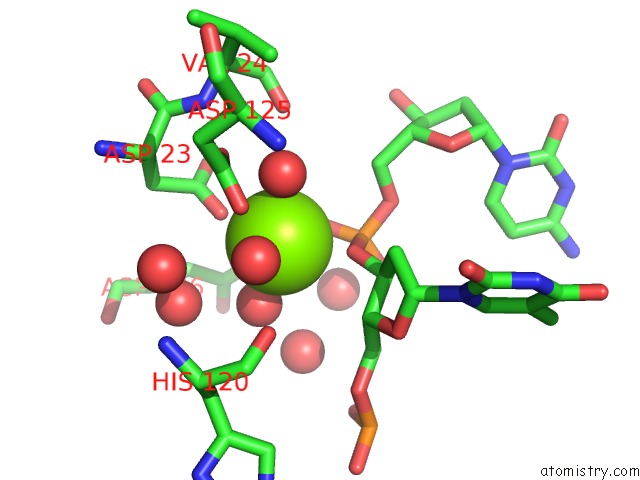
Mono view
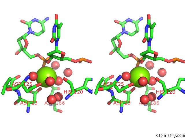
Stereo pair view
|
A full contact list of Magnesium with other atoms in the Mg binding
site number 3 of Crystal Structure of Rnase T in Complex with A Bluge Dna (Two Nucleotide Insertion Ct ) within 5.0Å range:
probe
|
atom
|
residue
|
distance (Å)
|
B
|
Occ
|
C:Mg101
b:19.4
occ:1.00
|
OD2
|
A:ASP23
|
2.3
|
27.3
|
1.0
|
O
|
A:HOH415
|
2.3
|
17.9
|
1.0
|
O
|
A:HOH424
|
2.3
|
20.6
|
1.0
|
O3'
|
C:DT17
|
2.4
|
13.6
|
1.0
|
O
|
A:HOH402
|
2.4
|
15.6
|
1.0
|
OP1
|
C:DC18
|
2.5
|
17.0
|
1.0
|
P
|
C:DC18
|
3.0
|
16.2
|
1.0
|
CG
|
A:ASP23
|
3.5
|
21.0
|
1.0
|
MG
|
A:MG301
|
3.5
|
26.3
|
1.0
|
C3'
|
C:DT17
|
3.6
|
11.5
|
1.0
|
O
|
A:HOH411
|
3.9
|
15.5
|
1.0
|
C4'
|
C:DT17
|
3.9
|
10.6
|
1.0
|
O5'
|
C:DC18
|
4.1
|
17.6
|
1.0
|
OD1
|
A:ASP23
|
4.1
|
30.4
|
1.0
|
OP2
|
C:DC18
|
4.2
|
16.5
|
1.0
|
C5'
|
C:DC18
|
4.2
|
14.9
|
1.0
|
O
|
A:HOH406
|
4.2
|
13.0
|
1.0
|
OD2
|
A:ASP125
|
4.2
|
16.1
|
1.0
|
C2'
|
C:DT17
|
4.3
|
12.8
|
1.0
|
O
|
A:VAL24
|
4.4
|
16.2
|
1.0
|
OD1
|
A:ASP125
|
4.5
|
17.2
|
1.0
|
CB
|
A:ASP23
|
4.6
|
16.5
|
1.0
|
C5'
|
C:DT17
|
4.6
|
14.5
|
1.0
|
O
|
C:HOH202
|
4.6
|
18.3
|
1.0
|
O
|
A:HIS120
|
4.7
|
15.5
|
1.0
|
OD2
|
A:ASP186
|
4.8
|
20.9
|
1.0
|
CG
|
A:ASP125
|
4.8
|
18.9
|
1.0
|
O
|
C:HOH209
|
4.9
|
27.1
|
1.0
|
|
Magnesium binding site 4 out
of 4 in 4kb1
Go back to
Magnesium Binding Sites List in 4kb1
Magnesium binding site 4 out
of 4 in the Crystal Structure of Rnase T in Complex with A Bluge Dna (Two Nucleotide Insertion Ct )
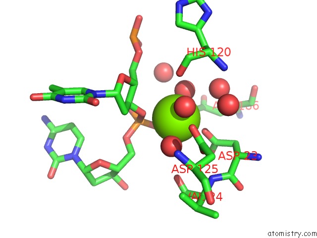
Mono view
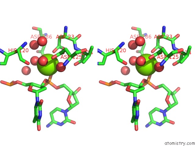
Stereo pair view
|
A full contact list of Magnesium with other atoms in the Mg binding
site number 4 of Crystal Structure of Rnase T in Complex with A Bluge Dna (Two Nucleotide Insertion Ct ) within 5.0Å range:
probe
|
atom
|
residue
|
distance (Å)
|
B
|
Occ
|
D:Mg101
b:17.4
occ:1.00
|
O
|
B:HOH426
|
2.2
|
19.2
|
1.0
|
O
|
B:HOH412
|
2.2
|
16.1
|
1.0
|
O
|
B:HOH424
|
2.4
|
19.3
|
1.0
|
OD1
|
B:ASP23
|
2.4
|
18.6
|
1.0
|
O3'
|
D:DT17
|
2.4
|
14.0
|
1.0
|
OP1
|
D:DC18
|
2.6
|
17.6
|
1.0
|
P
|
D:DC18
|
3.1
|
16.4
|
1.0
|
CG
|
B:ASP23
|
3.6
|
23.3
|
1.0
|
C3'
|
D:DT17
|
3.6
|
12.2
|
1.0
|
MG
|
B:MG301
|
3.8
|
30.8
|
1.0
|
O
|
B:HOH415
|
3.8
|
17.5
|
1.0
|
C4'
|
D:DT17
|
3.9
|
12.0
|
1.0
|
OD2
|
B:ASP125
|
4.0
|
17.9
|
1.0
|
C2'
|
D:DT17
|
4.1
|
15.4
|
1.0
|
C5'
|
D:DC18
|
4.1
|
11.9
|
1.0
|
O
|
B:HOH405
|
4.2
|
13.8
|
1.0
|
O5'
|
D:DC18
|
4.2
|
14.5
|
1.0
|
OD2
|
B:ASP23
|
4.2
|
24.7
|
1.0
|
OP2
|
D:DC18
|
4.3
|
19.0
|
1.0
|
O
|
B:VAL24
|
4.3
|
15.0
|
1.0
|
OD1
|
B:ASP125
|
4.4
|
16.6
|
1.0
|
OD2
|
B:ASP186
|
4.4
|
35.6
|
1.0
|
O
|
B:HIS120
|
4.6
|
13.5
|
1.0
|
CG
|
B:ASP125
|
4.6
|
17.7
|
1.0
|
O
|
D:HOH204
|
4.6
|
21.2
|
1.0
|
C5'
|
D:DT17
|
4.7
|
18.7
|
1.0
|
CB
|
B:ASP23
|
4.7
|
13.3
|
1.0
|
N
|
B:VAL24
|
5.0
|
12.3
|
1.0
|
|
Reference:
Y.Y.Hsiao,
W.H.Fang,
C.C.Lee,
Y.P.Chen,
H.S.Yuan.
Structural Insights Into Dna Repair By Rnase T--An Exonuclease Processing 3' End of Structured Dna in Repair Pathways. Plos Biol. V. 12 01803 2014.
ISSN: ISSN 1544-9173
PubMed: 24594808
DOI: 10.1371/JOURNAL.PBIO.1001803
Page generated: Sat Aug 17 03:32:15 2024
|