Magnesium »
PDB 4kg0-4knx »
4kg3 »
Magnesium in PDB 4kg3: Crystal Structure of Saccharomyces Cerevisiae DCP2 Nudix Domain in Complex with Mg (E153Q Mutation)
Protein crystallography data
The structure of Crystal Structure of Saccharomyces Cerevisiae DCP2 Nudix Domain in Complex with Mg (E153Q Mutation), PDB code: 4kg3
was solved by
R.A.Aglietti,
S.N.Floor,
J.D.Gross,
with X-Ray Crystallography technique. A brief refinement statistics is given in the table below:
| Resolution Low / High (Å) | 46.50 / 1.70 |
| Space group | C 1 2 1 |
| Cell size a, b, c (Å), α, β, γ (°) | 140.789, 49.270, 84.022, 90.00, 91.37, 90.00 |
| R / Rfree (%) | 17.7 / 21.3 |
Magnesium Binding Sites:
The binding sites of Magnesium atom in the Crystal Structure of Saccharomyces Cerevisiae DCP2 Nudix Domain in Complex with Mg (E153Q Mutation)
(pdb code 4kg3). This binding sites where shown within
5.0 Angstroms radius around Magnesium atom.
In total 3 binding sites of Magnesium where determined in the Crystal Structure of Saccharomyces Cerevisiae DCP2 Nudix Domain in Complex with Mg (E153Q Mutation), PDB code: 4kg3:
Jump to Magnesium binding site number: 1; 2; 3;
In total 3 binding sites of Magnesium where determined in the Crystal Structure of Saccharomyces Cerevisiae DCP2 Nudix Domain in Complex with Mg (E153Q Mutation), PDB code: 4kg3:
Jump to Magnesium binding site number: 1; 2; 3;
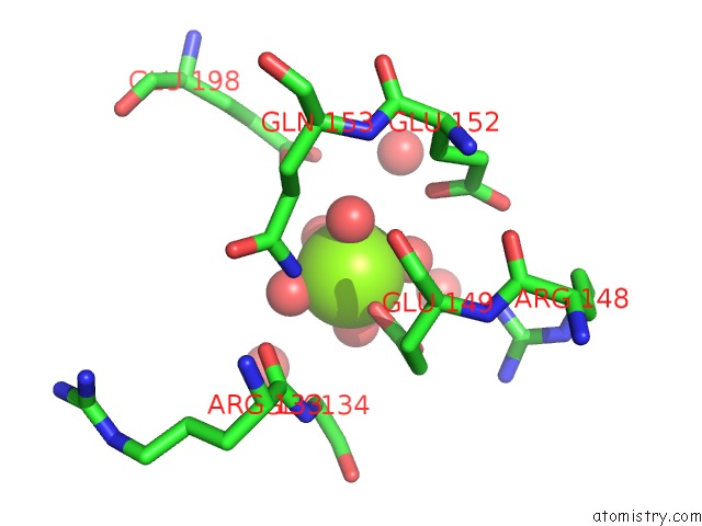
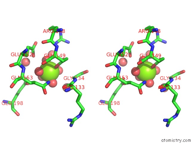
Magnesium binding site 1 out of 3 in 4kg3
Go back to
Magnesium binding site 1 out
of 3 in the Crystal Structure of Saccharomyces Cerevisiae DCP2 Nudix Domain in Complex with Mg (E153Q Mutation)
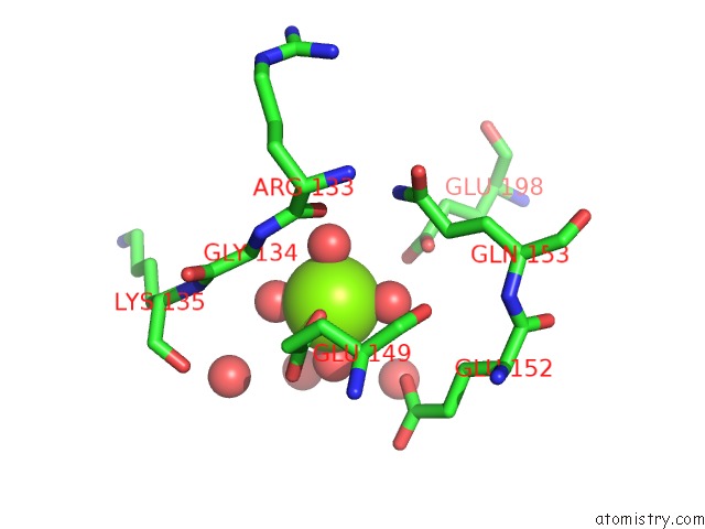
Mono view
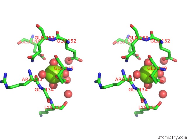
Stereo pair view
Mono view
Stereo pair view
A full contact list of Magnesium with other atoms in the Mg binding
site number 1 of Crystal Structure of Saccharomyces Cerevisiae DCP2 Nudix Domain in Complex with Mg (E153Q Mutation) within 5.0Å range:
|
Magnesium binding site 2 out of 3 in 4kg3
Go back to
Magnesium binding site 2 out
of 3 in the Crystal Structure of Saccharomyces Cerevisiae DCP2 Nudix Domain in Complex with Mg (E153Q Mutation)
Mono view
Stereo pair view
Mono view
Stereo pair view
A full contact list of Magnesium with other atoms in the Mg binding
site number 2 of Crystal Structure of Saccharomyces Cerevisiae DCP2 Nudix Domain in Complex with Mg (E153Q Mutation) within 5.0Å range:
|
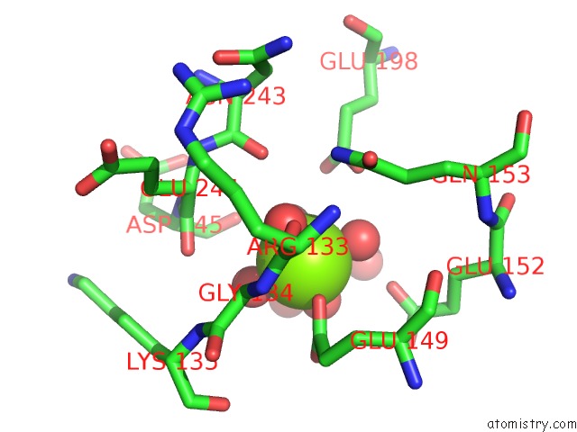
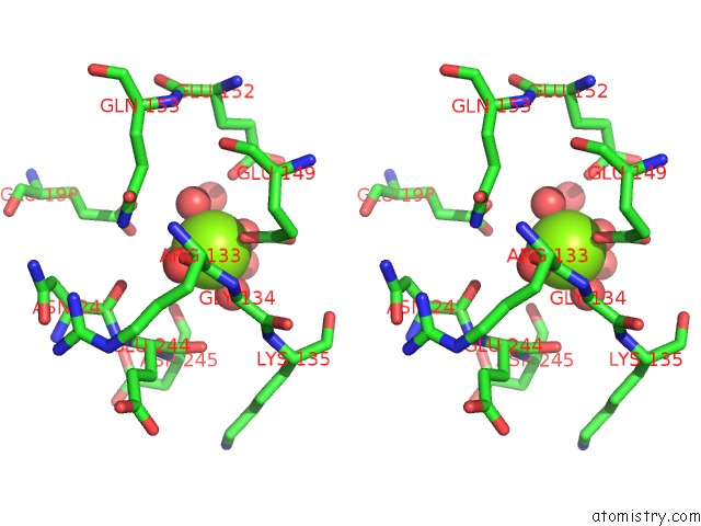
Magnesium binding site 3 out of 3 in 4kg3
Go back to
Magnesium binding site 3 out
of 3 in the Crystal Structure of Saccharomyces Cerevisiae DCP2 Nudix Domain in Complex with Mg (E153Q Mutation)
Mono view
Stereo pair view
Mono view
Stereo pair view
A full contact list of Magnesium with other atoms in the Mg binding
site number 3 of Crystal Structure of Saccharomyces Cerevisiae DCP2 Nudix Domain in Complex with Mg (E153Q Mutation) within 5.0Å range:
|
Reference:
R.A.Aglietti,
S.N.Floor,
C.L.Mcclendon,
M.P.Jacobson,
J.D.Gross.
Active Site Conformational Dynamics Are Coupled to Catalysis in the Mrna Decapping Enzyme DCP2. Structure V. 21 1571 2013.
ISSN: ISSN 0969-2126
PubMed: 23911090
DOI: 10.1016/J.STR.2013.06.021
Page generated: Sat Aug 17 03:36:04 2024
ISSN: ISSN 0969-2126
PubMed: 23911090
DOI: 10.1016/J.STR.2013.06.021
Last articles
Zn in 9MJ5Zn in 9HNW
Zn in 9G0L
Zn in 9FNE
Zn in 9DZN
Zn in 9E0I
Zn in 9D32
Zn in 9DAK
Zn in 8ZXC
Zn in 8ZUF