Magnesium »
PDB 4kg0-4knx »
4kgd »
Magnesium in PDB 4kgd: High-Resolution Crystal Structure of Pyruvate Oxidase From L. Plantarum in Complex with Phosphate
Enzymatic activity of High-Resolution Crystal Structure of Pyruvate Oxidase From L. Plantarum in Complex with Phosphate
All present enzymatic activity of High-Resolution Crystal Structure of Pyruvate Oxidase From L. Plantarum in Complex with Phosphate:
1.2.3.3;
1.2.3.3;
Protein crystallography data
The structure of High-Resolution Crystal Structure of Pyruvate Oxidase From L. Plantarum in Complex with Phosphate, PDB code: 4kgd
was solved by
P.Neumann,
K.Tittmann,
with X-Ray Crystallography technique. A brief refinement statistics is given in the table below:
| Resolution Low / High (Å) | 30.00 / 1.06 |
| Space group | C 2 2 21 |
| Cell size a, b, c (Å), α, β, γ (°) | 119.280, 154.160, 165.470, 90.00, 90.00, 90.00 |
| R / Rfree (%) | 12.7 / 15.1 |
Other elements in 4kgd:
The structure of High-Resolution Crystal Structure of Pyruvate Oxidase From L. Plantarum in Complex with Phosphate also contains other interesting chemical elements:
| Potassium | (K) | 1 atom |
Magnesium Binding Sites:
The binding sites of Magnesium atom in the High-Resolution Crystal Structure of Pyruvate Oxidase From L. Plantarum in Complex with Phosphate
(pdb code 4kgd). This binding sites where shown within
5.0 Angstroms radius around Magnesium atom.
In total 2 binding sites of Magnesium where determined in the High-Resolution Crystal Structure of Pyruvate Oxidase From L. Plantarum in Complex with Phosphate, PDB code: 4kgd:
Jump to Magnesium binding site number: 1; 2;
In total 2 binding sites of Magnesium where determined in the High-Resolution Crystal Structure of Pyruvate Oxidase From L. Plantarum in Complex with Phosphate, PDB code: 4kgd:
Jump to Magnesium binding site number: 1; 2;
Magnesium binding site 1 out of 2 in 4kgd
Go back to
Magnesium binding site 1 out
of 2 in the High-Resolution Crystal Structure of Pyruvate Oxidase From L. Plantarum in Complex with Phosphate
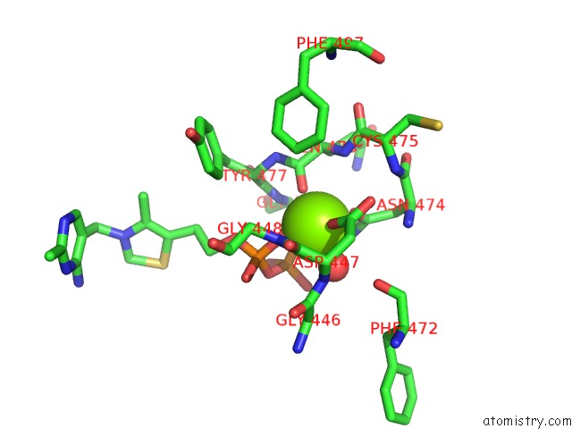
Mono view
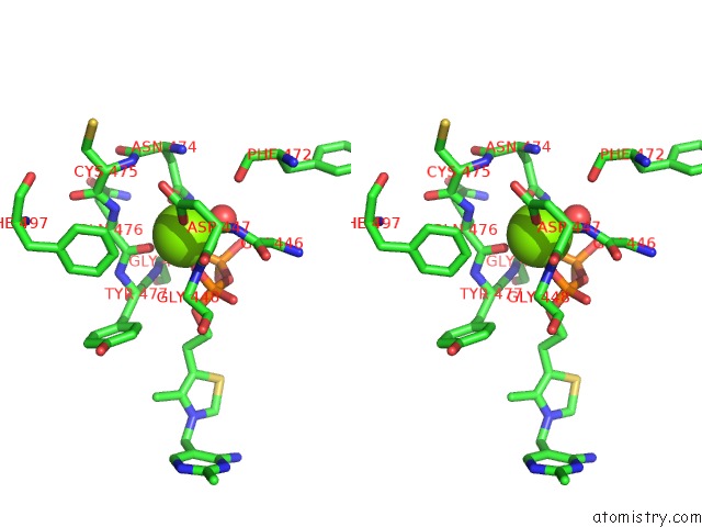
Stereo pair view
Mono view
Stereo pair view
A full contact list of Magnesium with other atoms in the Mg binding
site number 1 of High-Resolution Crystal Structure of Pyruvate Oxidase From L. Plantarum in Complex with Phosphate within 5.0Å range:
|
Magnesium binding site 2 out of 2 in 4kgd
Go back to
Magnesium binding site 2 out
of 2 in the High-Resolution Crystal Structure of Pyruvate Oxidase From L. Plantarum in Complex with Phosphate

Mono view

Stereo pair view
Mono view
Stereo pair view
A full contact list of Magnesium with other atoms in the Mg binding
site number 2 of High-Resolution Crystal Structure of Pyruvate Oxidase From L. Plantarum in Complex with Phosphate within 5.0Å range:
|
Reference:
D.Meyer,
P.Neumann,
R.Ficner,
K.Tittmann.
Observation of A Stable Carbene at the Active Site of A Thiamin Enzyme. Nat.Chem.Biol. V. 9 488 2013.
ISSN: ISSN 1552-4450
PubMed: 23748673
DOI: 10.1038/NCHEMBIO.1275
Page generated: Sat Aug 17 03:36:22 2024
ISSN: ISSN 1552-4450
PubMed: 23748673
DOI: 10.1038/NCHEMBIO.1275
Last articles
Zn in 9MJ5Zn in 9HNW
Zn in 9G0L
Zn in 9FNE
Zn in 9DZN
Zn in 9E0I
Zn in 9D32
Zn in 9DAK
Zn in 8ZXC
Zn in 8ZUF