Magnesium »
PDB 4ko8-4kvw »
4krc »
Magnesium in PDB 4krc: Crystal Structure of PHO85-PCL10-Atp-Gamma-S Complex
Enzymatic activity of Crystal Structure of PHO85-PCL10-Atp-Gamma-S Complex
All present enzymatic activity of Crystal Structure of PHO85-PCL10-Atp-Gamma-S Complex:
2.7.11.22;
2.7.11.22;
Protein crystallography data
The structure of Crystal Structure of PHO85-PCL10-Atp-Gamma-S Complex, PDB code: 4krc
was solved by
F.A.Quiocho,
F.Zheng,
with X-Ray Crystallography technique. A brief refinement statistics is given in the table below:
| Resolution Low / High (Å) | 39.44 / 2.60 |
| Space group | P 1 21 1 |
| Cell size a, b, c (Å), α, β, γ (°) | 50.145, 65.378, 80.254, 90.00, 100.61, 90.00 |
| R / Rfree (%) | 20.4 / 27 |
Magnesium Binding Sites:
The binding sites of Magnesium atom in the Crystal Structure of PHO85-PCL10-Atp-Gamma-S Complex
(pdb code 4krc). This binding sites where shown within
5.0 Angstroms radius around Magnesium atom.
In total only one binding site of Magnesium was determined in the Crystal Structure of PHO85-PCL10-Atp-Gamma-S Complex, PDB code: 4krc:
In total only one binding site of Magnesium was determined in the Crystal Structure of PHO85-PCL10-Atp-Gamma-S Complex, PDB code: 4krc:
Magnesium binding site 1 out of 1 in 4krc
Go back to
Magnesium binding site 1 out
of 1 in the Crystal Structure of PHO85-PCL10-Atp-Gamma-S Complex
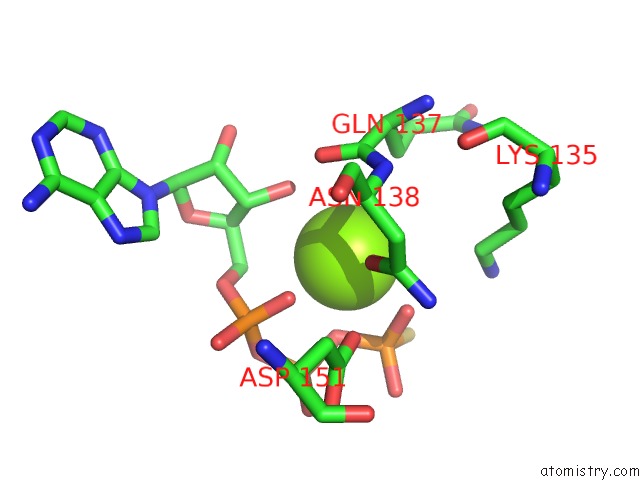
Mono view
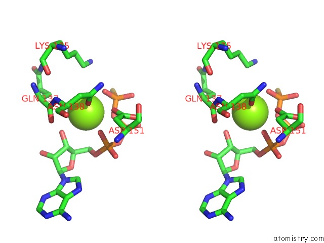
Stereo pair view
Mono view
Stereo pair view
A full contact list of Magnesium with other atoms in the Mg binding
site number 1 of Crystal Structure of PHO85-PCL10-Atp-Gamma-S Complex within 5.0Å range:
|
Reference:
F.Zheng,
F.A.Quiocho.
New Structural Insights Into Phosphorylation-Free Mechanism For Full Cyclin-Dependent Kinase (Cdk)-Cyclin Activity and Substrate Recognition. J.Biol.Chem. V. 288 30682 2013.
ISSN: ISSN 0021-9258
PubMed: 24022486
DOI: 10.1074/JBC.M113.502773
Page generated: Sat Aug 17 03:56:41 2024
ISSN: ISSN 0021-9258
PubMed: 24022486
DOI: 10.1074/JBC.M113.502773
Last articles
Zn in 9MJ5Zn in 9HNW
Zn in 9G0L
Zn in 9FNE
Zn in 9DZN
Zn in 9E0I
Zn in 9D32
Zn in 9DAK
Zn in 8ZXC
Zn in 8ZUF