Magnesium »
PDB 4ko8-4kvw »
4ktt »
Magnesium in PDB 4ktt: Structural Insights of Mat Enzymes: MATA2B Complexed with Sam
Enzymatic activity of Structural Insights of Mat Enzymes: MATA2B Complexed with Sam
All present enzymatic activity of Structural Insights of Mat Enzymes: MATA2B Complexed with Sam:
2.5.1.6;
2.5.1.6;
Protein crystallography data
The structure of Structural Insights of Mat Enzymes: MATA2B Complexed with Sam, PDB code: 4ktt
was solved by
B.Murray,
S.V.Antonyuk,
A.Marina,
S.C.Lu,
J.M.Mato,
S.S.Hasnain,
A.L.Rojas,
with X-Ray Crystallography technique. A brief refinement statistics is given in the table below:
| Resolution Low / High (Å) | 47.41 / 2.59 |
| Space group | P 21 21 21 |
| Cell size a, b, c (Å), α, β, γ (°) | 72.442, 115.723, 298.455, 90.00, 90.00, 90.00 |
| R / Rfree (%) | 21.8 / 27.9 |
Magnesium Binding Sites:
The binding sites of Magnesium atom in the Structural Insights of Mat Enzymes: MATA2B Complexed with Sam
(pdb code 4ktt). This binding sites where shown within
5.0 Angstroms radius around Magnesium atom.
In total only one binding site of Magnesium was determined in the Structural Insights of Mat Enzymes: MATA2B Complexed with Sam, PDB code: 4ktt:
In total only one binding site of Magnesium was determined in the Structural Insights of Mat Enzymes: MATA2B Complexed with Sam, PDB code: 4ktt:
Magnesium binding site 1 out of 1 in 4ktt
Go back to
Magnesium binding site 1 out
of 1 in the Structural Insights of Mat Enzymes: MATA2B Complexed with Sam
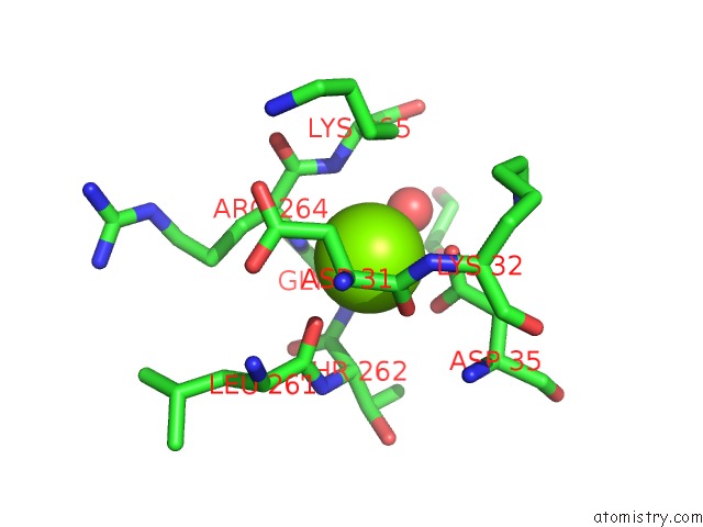
Mono view
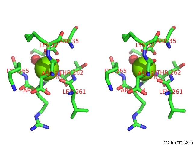
Stereo pair view
Mono view
Stereo pair view
A full contact list of Magnesium with other atoms in the Mg binding
site number 1 of Structural Insights of Mat Enzymes: MATA2B Complexed with Sam within 5.0Å range:
|
Reference:
B.Murray,
S.V.Antonyuk,
A.Marina,
S.M.Van Liempd,
S.C.Lu,
J.M.Mato,
S.S.Hasnain,
A.L.Rojas.
Structure and Function Study of the Complex That Synthesizes S-Adenosylmethionine. Iucrj V. 1 240 2014.
ISSN: ESSN 2052-2525
PubMed: 25075345
DOI: 10.1107/S2052252514012585
Page generated: Sat Aug 17 03:57:35 2024
ISSN: ESSN 2052-2525
PubMed: 25075345
DOI: 10.1107/S2052252514012585
Last articles
Zn in 9MJ5Zn in 9HNW
Zn in 9G0L
Zn in 9FNE
Zn in 9DZN
Zn in 9E0I
Zn in 9D32
Zn in 9DAK
Zn in 8ZXC
Zn in 8ZUF